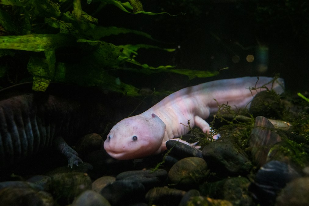
In the summer of 2011, in a basement lab at Tufts University in Medford, Massachusetts, a developmental biologist named Michael Levin was running a series of experiments that had nothing to do with limbs. His team was studying how planarian flatworms know how many heads to grow. But the axolotls kept sitting in tanks down the hall, doing something no other vertebrate on the planet can do: rebuilding an entire arm, bone, muscle, nerve, blood vessel, and skin, correctly proportioned, from a stump. Cut off an axolotl’s forelimb at the wrist and it grows back a wrist. Cut it at the shoulder and it grows back a full arm. The animal does not seem to guess. It seems to remember.
That distinction, guessing versus remembering, sits at the center of one of the strangest open questions in developmental biology. Salamanders, of which the axolotl (Ambystoma mexicanum) is the most studied example, do not regenerate limbs the way a scar heals over a wound. They regrow the specific missing anatomy, at the specific scale it was lost, using a positional memory that survives amputation, survives the death of the very cells that held it, and somehow re-writes itself into a fresh population of regenerating cells within days. For decades the leading explanation was purely chemical: gradients of retinoic acid, gradients of FGF and Wnt signaling proteins, a soup of diffusible molecules that pattern the new tissue the way they pattern an embryo. That explanation is not wrong. It is incomplete. Levin’s lab, and a small cohort of researchers working alongside him, have spent two decades building the case that axolotls also store and transmit positional information as bioelectric signal: voltage patterns held across the membranes of cells, patterns that persist and propagate independent of any single gene or protein.
The Blastema Reads a Voltage Map Before It Reads Any Gene
When an axolotl loses a limb, the wound does not simply scab over. Within hours, epidermal cells migrate across the stump and form a specialized cap. Beneath that cap, over the following days, mature cells at the injury site dedifferentiate: muscle fibers, bone cells, and fibroblasts lose their specialized identity and revert to a proliferative, stem-like state. This mass of dedifferentiated cells is called the blastema, and it is the structure that will rebuild the limb. The open question that obsessed researchers for most of the twentieth century was how the blastema knows what to build. A blastema formed at the wrist produces a hand. A blastema formed at the shoulder produces an entire arm, elbow included. The cells in both blastemas look identical under a microscope. Something invisible to histology is telling them where they are.
That something includes an electrical field. Salamander limb stumps generate a measurable current, first documented by Richard Borgens in the 1970s using vibrating probes sensitive enough to detect ion flow at the wound surface. Sodium ions rush into the stump and potassium and chloride flow out, driven by ion channels and pumps embedded in the epidermal cells covering the wound. This current is not a side effect of injury. It is a signal. Blocking the current pharmacologically, or reversing its polarity with an external electrode, changes how the blastema forms and, in some experimental conditions, disrupts regeneration outright. The wound current establishes a voltage gradient across the stump tissue, and cells within that tissue carry their own resting membrane potential, the voltage difference between the inside and outside of a cell maintained by ion channels and pumps. Depolarized cells sit at one part of the voltage map. Hyperpolarized cells sit at another. Levin’s work, building on earlier findings from Borgens and from Kenneth Robinson at Purdue, argues that this map functions as a coordinate system the blastema reads before it commits to any specific gene expression program.
The blastema does not build blindly. It reads a voltage map before it reads a single gene.
The clearest demonstration came from work on limb identity rather than limb presence. In classic experiments dating back to the mid-twentieth century and extended by Levin’s group with modern molecular tools, researchers found that manipulating the ion channels responsible for the resting potential in blastema cells could change the pattern of the regenerated structure, in some cases producing malformed or duplicated elements, without altering the underlying DNA sequence at all. The information governing the outcome lived in the electrical state of the tissue, not in a mutation.
Ion Channels, Not Neurons, Do Most of the Talking
It is tempting to assume that bioelectric signaling in regeneration must run through nerves, since nerves are the tissue most people associate with electricity in the body. Nerves matter here. Denervated axolotl limbs regenerate poorly or not at all, and the nerve-dependent factors involved, including a signaling protein identified by Anoop Kumar and Jeremy Brockes at University College London in 2005 called nAG, are essential for blastema growth. But the bioelectric patterning Levin’s lab describes operates largely outside neurons, in the ordinary somatic cells of skin, muscle, and connective tissue. Every cell in the body, not just neurons, maintains a resting membrane potential using ion channels and pumps, most notably the sodium potassium ATPase pump and a range of voltage gated and ligand gated channels. In an axolotl blastema, these non-neural cells electrically couple to one another through gap junctions, protein channels that let ions and small molecules pass directly from one cell’s cytoplasm into its neighbor’s. That coupling turns a field of individual cells into something closer to a single distributed electrical circuit, capable of holding a stable pattern across thousands of cells simultaneously.
This is the mechanism that lets the memory outlast the original cells. A gene expressed in one cell dies with that cell. A voltage pattern maintained across a gap junction coupled network can, in principle, be reconstituted by neighboring cells even after the originating cells are gone, because the pattern is a property of the network’s connectivity and channel activity, not of any single cell’s contents. Levin has described this as analogous to how a pattern can persist in a flock of birds even as individual birds enter and leave the flock. The formation is the information. Disrupt the gap junctions with a pharmacological blocker such as octanol or heptanol, and blastema patterning goes wrong even when every gene involved is intact and every growth factor is present at normal concentration. The electrical connectivity itself was carrying instructions that biochemistry alone could not supply.
A gene dies with its cell. A voltage pattern held in a coupled network can outlive the cells that first held it.
Dany Adams, working with Levin’s group, used voltage sensitive fluorescent dyes to visualize this directly in regenerating Xenopus tadpole tails, a related regenerative system studied alongside axolotls because it is faster to image and easier to manipulate. The dyes change brightness in proportion to membrane voltage, turning an invisible electrical pattern into a movie. What that movie showed was a wave of depolarization sweeping through the wound site hours before any visible regrowth, followed by the stabilization of distinct high and low voltage domains that mapped onto where the regenerating structure would eventually take shape. The electrical pattern was not a byproduct of growth. It arrived first.
Rewriting the Map Can Rewrite the Limb
The strongest evidence for bioelectric memory is not descriptive, it is interventional. If voltage patterns are merely correlated with regeneration outcomes, changing the voltage should do nothing. If voltage patterns actually encode instructions, changing the voltage should change the outcome, even without touching genes directly. Levin’s lab and collaborators have run this experiment repeatedly, most dramatically not in axolotls but in the related tadpole system, where they used ion channel drugs, including ivermectin and other channel modulating compounds, to force a patch of tissue on a tadpole’s flank into a depolarization pattern that normally appears only at the site of an eye. The result, published in 2007, was a fully formed, functional eye, complete with lens and retina, growing on the flank, wired into the tadpole’s optic nerve, in a location that never received any eye specific genetic signal. The instruction to build an eye was not delivered by a gene. It was delivered by a voltage state that the surrounding tissue interpreted using its own default developmental program.
In axolotls specifically, similar logic explains one of the oldest puzzles in the field: why blastemas at different amputation levels produce different amounts of limb. Researchers including Susan Bryant and David Gardiner at UC Irvine spent decades mapping what they called positional memory using grafting experiments, moving blastema tissue from one part of a limb to another and watching the graft fill in exactly the segment appropriate to its original position, regardless of where it was transplanted. A wrist level blastema grafted onto a shoulder stump still tries to build a wrist’s worth of structure, then relies on intercalation, a process where the surrounding tissue fills in the missing middle segments to reconcile the mismatch. That positional memory has to be stored somewhere physically durable enough to survive both amputation and transplantation. Levin’s bioelectric framework offers the first mechanism that plausibly explains how such memory could be stored in ordinary tissue without requiring a permanent, hardwired genetic map for every possible amputation level, because a voltage state can be re-established, referenced, and even overwritten far more flexibly than a fixed genetic circuit could be.
| Mechanism | Key Researcher(s) | What It Explains |
|---|---|---|
| Wound epithelium current | Richard Borgens, 1970s | Initial ion flow that triggers blastema formation |
| Gap junction coupling | Michael Levin, Tufts University | How voltage patterns span many cells as one network |
| Nerve dependent nAG signaling | Anoop Kumar, Jeremy Brockes, UCL, 2005 | Why denervated limbs fail to regenerate |
| Positional memory grafting | Susan Bryant, David Gardiner, UC Irvine | Why blastemas rebuild the correct missing segment |
| Ectopic eye induction via depolarization | Levin lab, Xenopus, 2007 | Voltage alone can trigger organ specific programs |
Why Mammals Mostly Lost This and What That Costs Us
Humans possess nearly all the same molecular parts axolotls use. Human cells have resting membrane potentials, gap junctions, ion channels, and the same core developmental gene families. Human fingertips, in infants and occasionally in young children, can even regenerate a distal segment after amputation, a limited echo of what axolotls do routinely across an entire limb. What humans lack is the whole system operating together at scale: a wound epidermis that behaves like the specialized apical cap of a salamander, a durable multicellular bioelectric pattern that survives injury, and a population of cells willing to dedifferentiate broadly rather than simply scarring over. Mammalian wound healing evolved to prioritize speed and infection control, sealing a wound with fibrous scar tissue within days. That scar, useful for survival in a world full of pathogens, is also a wall. It blocks the kind of large scale cellular dedifferentiation and bioelectric network formation that axolotl wounds permit.
This is why Levin’s research program extends past axolotls into frogs, planarians, and eventually mammalian tissue culture and cancer models, where his lab has shown that disrupting normal bioelectric states in otherwise healthy frog cells can induce tumor like growth, and that restoring normal voltage patterns can, in some cases, normalize cells that carry cancer driving mutations. If voltage state can override genetic instruction in one direction, inducing an eye where none was coded, it plausibly can be steered in the other direction too, toward suppressing aberrant growth or toward instructing a wound to build a structure instead of a scar. No lab has produced a regenerating human limb, and nobody studying this seriously claims that is imminent. What the axolotl demonstrates, cleanly and repeatedly, is that a vertebrate body already contains a second information layer beyond DNA, one that specifies not what a cell is capable of building, but which of its capabilities to build right now, at this location, at this scale.
The axolotl’s limb is not rebuilt from a genetic blueprint alone. It is rebuilt from a blueprint plus an address, and the address is written in volts. Somewhere in the gap between a stump and a fully formed shoulder, wrist, and set of fingers, a field of ordinary cells briefly becomes a kind of distributed nervous system for a problem that has nothing to do with thought: not what should this cell become, but where, exactly, does this cell think it is standing.
Credit: Mattias Banguese on Unsplash