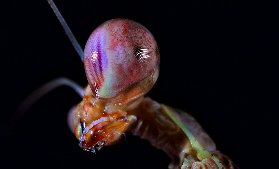
In 2014, a team led by Hanne Thoen and Justin Marshall at the Queensland Brain Institute published a finding in Science that embarrassed a decade of assumptions about the mantis shrimp’s famous eye. The animal has somewhere between 12 and 16 classes of photoreceptor, more than double what humans carry, and researchers had long assumed that meant mantis shrimp could see color with extraordinary precision. Thoen’s behavioral tests showed the opposite. Mantis shrimp are worse than humans at telling similar wavelengths apart. The extra receptors weren’t buying finer color vision. They were buying something else entirely, a different way of turning light into electrical signal that skips a step most animal brains can’t skip.
That finding sat mostly in the realm of vision science for a decade. It’s now migrating into bioelectronic medicine, because the same molecular trick that makes mantis shrimp eyes strange is showing up in opsins being engineered to restore sight in blind human retinas. Sonja Kleinlogel’s lab at the University of Bern has spent the past several years isolating and re-purposing the light-sensitive proteins from stomatopod eyes, and the company she co-founded, Arctos Medical, is building an optogenetic therapy around them. The mantis shrimp’s weird eye turns out to be a better starting material for retinal gene therapy than the algae proteins most optogenetics labs have relied on since the mid-2000s.
Sixteen Receptors, Worse Color Vision Than You’d Expect
Most animals with color vision, humans included, do it through opponent processing. The brain compares the output of a small number of receptor types, say a signal from a “red” cone against a signal from a “green” cone, and the difference between those two electrical readings is what gets interpreted as hue. It’s an efficient system because you only need two or three receptor classes to cover a wide range of colors, but it demands real neural computation. Comparison happens downstream, in retinal ganglion cells and visual cortex, using circuitry that has to be wired with some precision.
Mantis shrimp don’t appear to do this. Thoen’s experiments suggested that instead of comparing receptors against each other, the animal scans a scene with its eye’s midband region, sweeping different photoreceptor rows across a point of interest and reading each one out close to sequentially. Color identification becomes closer to checking which of sixteen pre-tuned bins lit up rather than computing a difference between two continuous values. It requires more receptor types than opponent processing does, but it requires far less neural math to interpret them.
More light sensors didn’t buy the shrimp better color vision. It bought a shortcut around comparison.
That distinction matters for anyone thinking about bioelectric systems generally, not just vision. It’s a real example of a nervous system trading receptor diversity for processing simplicity, pushing information encoding out to the periphery, into the sensor itself, instead of paying for it later in synapses and spikes. Marshall’s group has described the mantis shrimp visual system for years as running more like a scanner than a camera. The bioelectric signal coming out of each photoreceptor row barely needs interpretation once it reaches the brain, because the identity of which row fired already carries most of the meaning.
The Molecule Doing the Work Is a Switch, Not Just a Sensor
The part of this story with direct medical relevance sits one level below behavior, in the opsin proteins themselves. Human rod and cone opsins are monostable. Light flips the molecule into an active shape, and a separate biochemical cycle, the visual cycle, has to regenerate it back to a resting state before it can fire again. That regeneration step is slow and metabolically expensive, and it’s one reason human eyes struggle in rapidly changing light and why a bleached retina takes real time to recover.
Several of the opsins in the mantis shrimp midband are bistable. One wavelength of light pushes the molecule into an active, signal-generating state. A different wavelength pushes it right back to resting, without needing the cell’s separate regeneration machinery at all. The opsin functions as a two-way optical switch built directly into the membrane’s bioelectric machinery, on and off, no chemical cleanup crew required in between. Kleinlogel’s lab named this family of proteins MerMAID, isolated from mantis shrimp middle-wavelength photoreceptors, and published the core characterization in Science Advances in 2020.
The eye did the work the brain usually does, and it did it with a molecule that resets itself.
For basic vision science this was an interesting structural quirk. For anyone trying to build a light-activated therapy, it’s close to the ideal property list. A retinal cell engineered to express a bistable opsin doesn’t wait around for slow chemical regeneration between light pulses, and it doesn’t require the constant, high-intensity illumination that faster-desensitizing algae opsins need to stay useful. That combination directly addresses two of the practical failure points that have slowed optogenetic vision therapy since the field’s first human trials.
Turning a Shrimp’s Switch Into a Human Retinal Therapy
Optogenetic vision restoration works on a simple premise. In diseases like retinitis pigmentosa, the rods and cones that normally convert light into electrical signal die off, but the downstream retinal neurons, the bipolar and ganglion cells that would relay that signal to the brain, often survive for years without ever receiving input. Deliver a gene encoding a light-sensitive channel or receptor into those surviving cells with a viral vector, and you can make them light-sensitive on their own, bypassing the dead photoreceptors entirely.
The first generation of this approach used channelrhodopsins pulled from green algae, the same tool kit that built optogenetics as a neuroscience method in the first place. José-Alain Sahel and Botond Roska ran the therapy furthest into humans with GenSight Biologics’ PIONEER trial, publishing in Nature Medicine in 2021 that a blind retinitis pigmentosa patient regained enough light perception, using ChrimsonR paired with light-stimulating goggles, to locate and touch objects on a table. It was a genuine milestone and also a clear demonstration of the limits: the therapy required goggles delivering intense, specifically timed amber light, and the restored vision was crude, more like detecting shapes and motion than seeing a scene.
MerMAID opsins are being developed to push past that ceiling. Because the switch is bistable, a therapy built on it can, in principle, be driven by ordinary ambient light rather than requiring a dedicated high-intensity light source strapped to the patient’s face. Arctos Medical’s preclinical work is aimed at retinitis pigmentosa and other photoreceptor degenerations, using the same gene therapy delivery logic as the algae-based approaches, a viral vector carrying the opsin gene into surviving bipolar cells, but with a light-sensing molecule that behaves more like the eye’s own equipment.
| Opsin | Source organism | Key group | Switching behavior | Status |
|---|---|---|---|---|
| Channelrhodopsin-2 | Chlamydomonas reinhardtii (algae) | Foundational optogenetics tool, multiple labs | Monostable, requires continuous illumination | First human retinal trials, mid-2010s onward |
| ChrimsonR | Chlamydomonas noctigama (algae) | Sahel/Roska, GenSight Biologics PIONEER trial | Monostable, amber light activated with goggles | Partial vision restoration reported, 2021 |
| MerMAID family | Mantis shrimp (stomatopod) photoreceptors | Sonja Kleinlogel, University of Bern / Arctos Medical | Bistable, switched by two wavelengths | Preclinical development |
Where This Runs Into a Wall
Here’s the honest catch, and it comes straight from the same behavioral data that started this story. Mantis shrimp have more photoreceptor types than almost any animal on Earth and still test worse than humans at fine color discrimination. That’s a warning against a specific, seductive assumption: that adding more light-sensitive channel types to a retina, or engineering flashier switching kinetics, automatically buys better vision. Marshall’s work shows the opposite can be true. The mantis shrimp traded discrimination for speed and simplicity, and it works for an animal that needs to react fast underwater, not for one that needs to read a menu.
Applied to retinal therapy, that means a bistable opsin solves a real engineering problem, the light intensity and refresh-rate limits that plague algae-derived channels, without automatically solving the harder problem of restoring anything close to natural color perception or spatial resolution. The vision these therapies produce, at least in the current generation, remains coarse. Patients in the ChrimsonR trial described perceiving light and motion, not scenes. There’s no published human data yet on MerMAID-based therapy, and going from a mouse retina expressing a mantis shrimp opsin to a functioning treatment in a person carries the usual multi-year gap between preclinical promise and an approved product. Bistability fixes a hardware problem. It doesn’t rewrite the software the visual cortex uses to interpret a sparse, artificially generated signal.
A photoreceptor that resets itself is a photoreceptor you can trust in a blind eye. It still isn’t a cure.
The gap between “the switch works” and “the patient sees” is where most optogenetic vision programs have spent the last ten years, and there’s no evidence a shrimp-derived opsin gets to skip that part.
What a Shrimp’s Eye Says About Bioelectric Design Generally
Step back from the retina and the mantis shrimp case makes a broader point about how living systems handle information at the point where physics turns into voltage. You can build sophistication into the sensor, the way stomatopods packed sixteen receptor types and a self-resetting molecular switch into their eyes, or you can build it into the circuit that reads the sensor out, the way human opponent-processing vision does with three cone types and a lot of downstream comparison. Neither strategy is objectively better. They’re different bioelectric budgets, spending complexity in different places depending on what the organism actually needs to survive.
That framing is useful for anyone building neural interfaces or sensory prosthetics beyond vision, because it’s a live design choice every time: put the intelligence in the electrode and the transducer, or put it in the software decoding the signal afterward. Bistable mantis shrimp opsins are a case where nature already solved the transducer-side version of that problem, tens of millions of years before anyone needed it for a gene therapy. Kleinlogel’s group didn’t invent the switch. They found it sitting in a shrimp’s eye and figured out how to put it into a human retinal cell instead.
The open question is whether that borrowed molecular trick ends up mattering for patients the way ChrimsonR already has, or whether it stays a better-engineered tool without a correspondingly better clinical result. Mantis shrimp evolved their scanning, self-resetting visual system to survive on a reef, not to restore sight to a human retina, and there’s no guarantee the same properties that make the system efficient for a shrimp translate cleanly into better vision for someone with retinitis pigmentosa. The biology is elegant. Whether it clears a human trial is still an open bet.
Credit: Andrey Tikhonovskiy on Unsplash