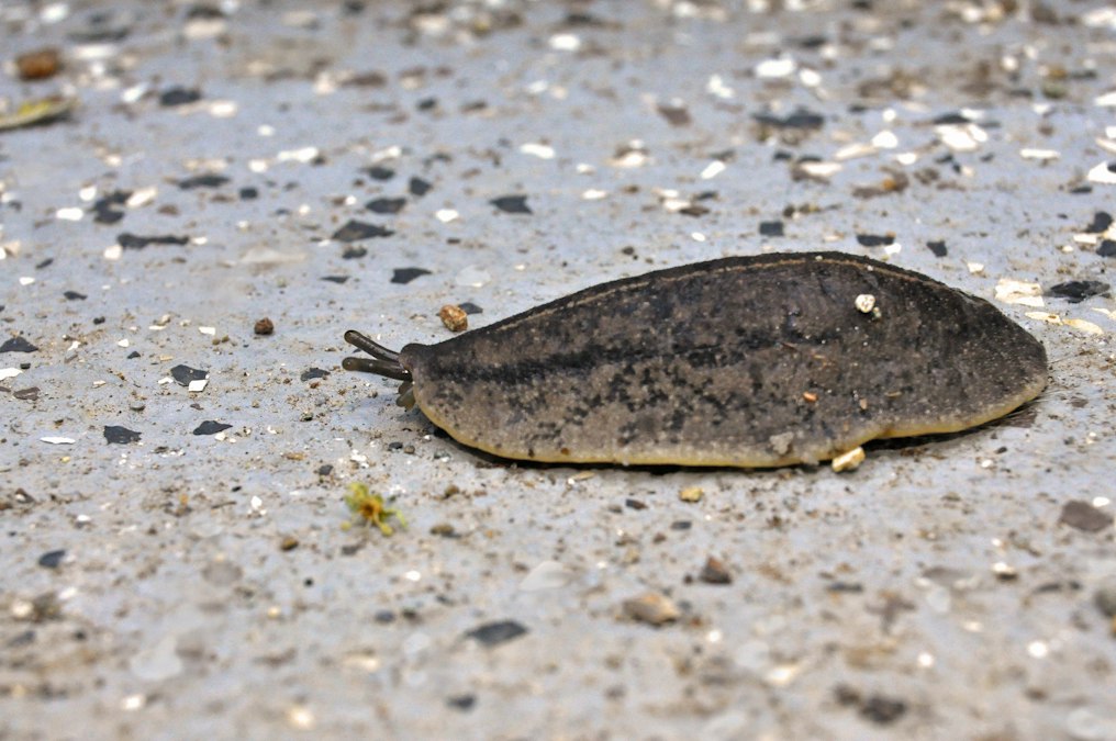
In 2013, Tal Shomrat and Michael Levin at Tufts University published a paper describing flatworms that would not stop growing two heads. The worms were planaria, the same brownish, cross-eyed creatures familiar from middle school biology for their ability to regenerate an entire body from a sliver of tissue. Shomrat and Levin cut the worms in half and briefly soaked the fragments in a drug called octanol, which blocks the channels that let cells pass electrical signals to their neighbors. No genes were edited. No DNA was touched. And yet a notable fraction of the treated fragments regenerated with a head at both ends, and kept doing so for months, through repeated amputations, long after the octanol had washed out of their tissue.
That persistence is the part worth sitting with. The worm’s genome never changed. What changed, and stayed changed, was a pattern of voltage across its cells, a kind of electrical layout that told the regenerating tissue where a head belongs. The genome supplied the parts. Something else supplied the blueprint.
Every cell in your body is sitting on a tiny battery
You’ve probably heard that neurons fire using electrical signals. What’s less widely known is that nearly every cell in the body, not just neurons, maintains a voltage difference across its outer membrane, usually somewhere between negative 10 and negative 90 millivolts relative to the fluid outside. This resting potential comes from pumps and channels that control which charged ions, mostly sodium, potassium, calcium, and chloride, sit inside the cell versus outside it. Neurons use rapid voltage spikes to send messages over long distances in milliseconds. Other cells use slower, steadier voltage states to signal something different: not “fire now,” but “this is what I am, and this is what I’m telling my neighbors to become.”
Those slower voltage states turn out to carry real instructions. Cells that are actively proliferating, including stem cells and, notably, cancer cells, tend to sit at a depolarized voltage, closer to zero. Cells that have finished differentiating into mature tissue tend to be more hyperpolarized, sitting at a more negative voltage. This isn’t just correlation. Researchers in Levin’s lab and others have shown that artificially shifting a cell’s voltage, using ion channel drugs or genetically encoded channels that respond to light, can push cells toward dividing or toward settling down, independent of what their genes alone would predict.
Cells share this information with each other through gap junctions, direct physical channels that connect the interior of one cell to the interior of its neighbor, letting ions and small molecules flow between them without ever touching the extracellular space. A field of connected cells behaves less like a crowd of individuals and more like a circuit board, with voltage patterns propagating across tissue the way current moves through wiring.
The genome supplied the parts. Something else supplied the blueprint.
The experiment that put an eye where an eye doesn’t belong
If voltage patterns were simply a readout of gene expression, a downstream effect with no independent power, none of this would matter much for how we think about development. The experiment that argued otherwise came out of Levin’s lab in 2012, published in the journal Development by Vaibhav Pai, Sherry Aw, and colleagues. They took Xenopus laevis frog embryos and forced cells in the gut, tissue that normally has nothing to do with vision, to express an ion channel that shifted their membrane voltage to match the depolarized state normally found in the embryonic eye field. A meaningful fraction of those embryos grew a well-formed eye, with lens and retinal tissue, on their gut.
No eye-specific transcription factor was switched on directly. The voltage state alone was enough to recruit the local gene networks needed to build an eye, in a location that had never been instructed to build one. That is the difference between voltage as a side effect and voltage as a cause. It means a tissue’s electrical state can function as an instruction a cell reads before it decides which genes to switch on, not just a side effect of genes that are already switched on.
Specificity doesn’t always come from where the tissue sits. Sometimes it comes from how it’s charged.
Wounds generate their own electric fields, and cells follow them
The clearest everyday example of bioelectric signaling doesn’t require a lab manipulation at all. Cut your skin, and the wound generates a measurable direct current electric field, typically on the order of tens to a couple hundred millivolts per millimeter, that persists for as long as the wound stays open. This happens because skin maintains an ion gradient across its outer layers, and breaking that layer short circuits the gradient, driving current to flow out of the wound and setting up a field across the surrounding tissue.
Colin McCaig at the University of Aberdeen and Richard Nuccitelli, working independently over decades, showed that skin cells migrate along these endogenous fields with striking directional precision, a phenomenon called galvanotaxis. Keratinocytes, the migrating skin cells that close a wound, orient and crawl toward the negative pole of the field, essentially following the exposed wound site the way a compass needle follows magnetic north. Block the field artificially, using drugs that interfere with the ion pumps generating it, and wound closure slows measurably. Apply an external field matching the body’s own polarity, and migration speeds up.
This is why electrical stimulation devices for chronic, slow-healing wounds, the kind seen in diabetic ulcers, aren’t a fringe idea. They’re an attempt to reinforce a signal the body already generates on its own but sometimes generates too weakly to finish the job.
Where this gets genuinely difficult
Here’s the honest limitation. Planarian flatworms and frog embryos are extraordinarily permissive systems for this kind of experiment. They tolerate dramatic developmental reprogramming, they regenerate readily, and their bioelectric networks are comparatively simple and well mapped after years of targeted study. A mouse, let alone a person, is a much harder case. Mammalian regeneration is sharply limited to begin with, scarring rather than rebuilding is the default response to most serious injuries, and the bioelectric maps that exist for mammalian tissue are far less complete than the maps Levin’s group and others have built for worms and frogs.
There’s also a real scientific debate about how much of this generalizes. Voltage shifts are clearly sufficient to trigger specific developmental programs in these model organisms under controlled conditions. Whether modulating voltage could reliably redirect healing or regeneration in a human limb, rather than just nudging migration speed at a wound edge, is a much bigger claim, and one that hasn’t been demonstrated. The field has had real, replicated results. It has not yet had a result that lets a mammal regrow something it couldn’t regrow before.
What the pattern looks like across systems
Laid side by side, the experiments share a logic even though the organisms and methods differ enormously.
| Mechanism | System Studied | Observed Effect | Key Lab |
|---|---|---|---|
| Gap junction blockade (octanol) | Planarian flatworms | Stable two-headed regeneration across repeated amputations | Levin lab, Tufts University |
| Forced ion channel expression | Xenopus embryo gut tissue | Ectopic, functional eye structures | Levin lab, Tufts University |
| Endogenous wound electric field | Mammalian and amphibian skin | Directed keratinocyte migration toward wound center | McCaig, University of Aberdeen; Nuccitelli, UC Davis |
In each case, the voltage pattern isn’t decoration. It’s doing work that would otherwise have to be done by, or would be impossible for, the genome alone acting on its own schedule.
What this actually buys regenerative medicine
The practical promise isn’t that doctors will soon dial a voltage knob and regrow a finger. It’s narrower and more useful than that. If voltage states are a layer of instruction that sits between gene expression and final tissue shape, then it becomes a layer you can intervene on without the difficulty of gene therapy, using existing, often already-approved ion channel drugs, or using targeted electrical stimulation delivered through a device. That’s a genuinely different lever than editing DNA, and it’s one reason bioelectricity has drawn attention from people working on wound care, birth defect research, and even early-stage cancer biology, where disrupted voltage states in tissue show up well before a tumor is large enough to find any other way.
The limiting factor isn’t the concept. It’s the map. Researchers can read and write voltage patterns in a flatworm or a frog embryo because decades of focused work went into characterizing exactly which channels sit where and what they do when nudged. No comparable map exists yet for human tissue at that resolution. Building one is slower, less glamorous work than any single striking experiment, and it’s the work that decides whether this becomes a treatment or stays a demonstration.
The flatworm that keeps regenerating two heads isn’t a curiosity. It’s a cell population that got handed new instructions through voltage alone and kept following them long after the original signal was gone. That persistence is the real finding: bioelectric pattern can outlast the event that set it, which means it functions less like a momentary nudge and more like a second, slower form of memory sitting alongside the genome. The open question isn’t whether that memory exists. It’s how much of it humans still have access to, and how much was lost somewhere between a frog that can regrow a tail and a species that mostly just scars.
Credit: Karl Callwood on Unsplash