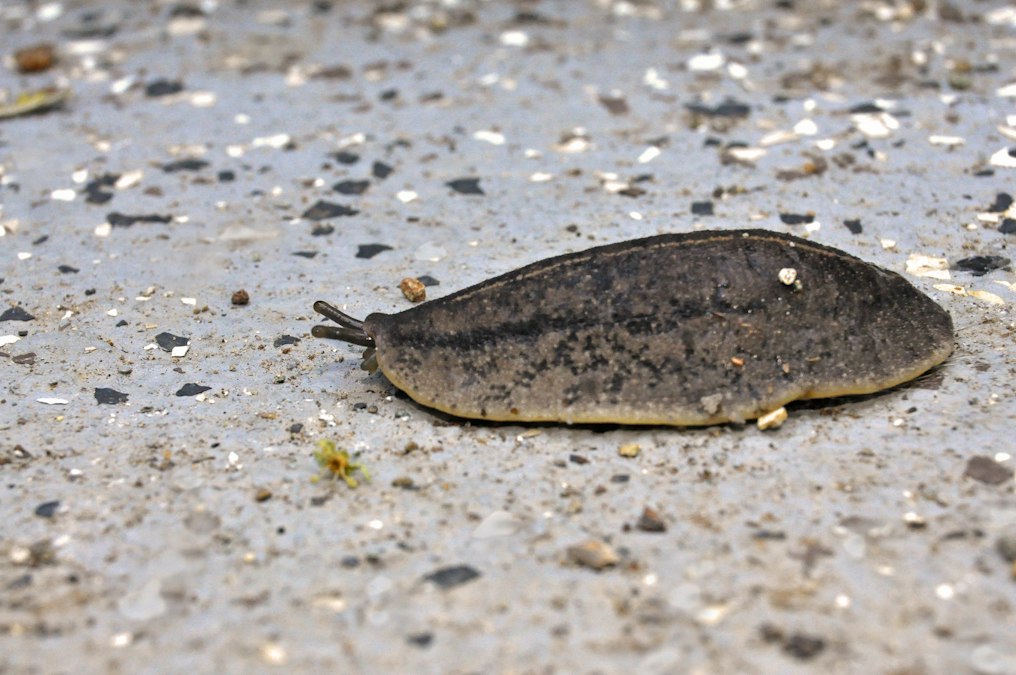
In 2011, a developmental biologist named Michael Levin, then building what would become the Allen Discovery Center at Tufts University, published a result that still unsettles people who hear it for the first time. His lab took flatworms called planaria, cut them in half, and before the animals regenerated, altered the voltage pattern across their cells using ion channel blocking drugs. No genes were edited. No DNA was touched. The worms grew back with two heads instead of one. Cut those two headed worms again, with no further intervention, and they kept regenerating as two headed animals indefinitely. The genome was identical to a normal planarian’s the entire time.
That result is the cleanest demonstration of a fact that is easy to state and hard to absorb: the instructions for where a body puts its parts are not stored only in DNA. They are stored, in part, in voltage. Every living cell holds an electrical charge across its outer membrane, a few tens of millivolts, maintained by pumps and channels working constantly. Tissues organize those individual charges into larger patterns, and those patterns carry information that genes read, not the other way around. This is the subject of bioelectricity, and it is one of the oldest ideas in biology that modern tools are only now letting researchers actually test.
Every Cell Runs a Voltage, All the Time, Whether You Notice or Not
A neuron firing is the most familiar example of bioelectricity, a rapid voltage spike that resets itself in milliseconds. But every cell in your body, not just neurons, maintains a steady resting voltage across its membrane, typically between negative 20 and negative 90 millivolts relative to the outside. That voltage exists because pumps embedded in the membrane, the sodium potassium ATPase chief among them, spend enormous amounts of cellular energy shoving ions against their concentration gradients. Roughly a third of your resting metabolic energy goes to running these pumps. You are, in a literal sense, a battery that costs calories to keep charged.
What makes this voltage more than a cellular housekeeping detail is that cells are not electrically isolated from their neighbors. Many tissues connect through gap junctions, channel proteins called connexins that let ions and small molecules flow directly from one cell’s interior to the next. A sheet of connected cells behaves like a single electrical network, and the voltage pattern across that network, the sum of thousands of individual resting potentials, can encode shape information the way a stencil encodes an outline. Change the voltage pattern and you change what the tissue builds, independent of changing which genes are present.
You are a battery that costs calories to keep charged.
This was not a new idea in 2011. Harold Saxton Burr, a Yale anatomist working from the 1930s through the 1950s, spent decades measuring what he called electro-dynamic fields around developing salamander eggs and other organisms, and claimed the voltage pattern predicted the future axis of growth before any visible structure appeared. Burr’s instruments were crude by today’s standards and his broader theorizing drifted into territory mainstream biology never accepted, but his core observation, that measurable electrical fields precede and track anatomical development, held up. Lionel Jaffe and Kenneth Robinson, working at Purdue in the 1970s, built a vibrating probe sensitive enough to measure the tiny ionic currents flowing through embryos and wounds, giving Burr’s old observation an actual number attached to it for the first time.
The Flatworm Experiment Nobody Expected to Work
Levin’s two headed planaria matter because they separate two things that are normally tangled together in biology: the genome, which is fixed, and the bioelectric state, which is not. The worms’ DNA never specified “grow two heads.” Their voltage pattern did, and the downstream gene expression machinery simply executed whatever the voltage pattern told it to build. Later experiments from the same lab pushed this further. Expressing specific ion channels to shift the resting potential of cells in the gut or tail region of a frog tadpole, work done in Xenopus laevis, caused those cells to grow an eye, complete with lens and retina, in a location that had never received any eye specifying genetic signal. The cells had the genes to build an eye. They needed the voltage cue telling them to use them.
The worms kept regenerating as two headed animals, with no genetic edit involved at all.
The term Levin’s group uses for this is a bioelectric code, a layer of pattern information sitting above gene regulatory networks, readable and in some cases rewritable with drugs that open or close specific ion channels, without touching a single base pair of DNA. It reframes a question biologists have asked since Hans Driesch’s sea urchin experiments in the 1890s, the question of how a collection of genetically identical cells knows to become a head here and a tail there. Genes supply the parts list. Something else decides where the parts go, and voltage is a leading answer for what that something else is.
Your Skin Starts Repairing Itself With a Current, Not a Chemical
Bioelectric signaling is not confined to exotic flatworm experiments. It happens in every wound you have ever gotten. Intact skin maintains a voltage gradient across its layers, driven by ion pumps in epithelial cells, and when that skin is broken, the gradient discharges at the wound site and generates a local electric field, typically on the order of 100 to 200 millivolts per millimeter, that persists for as long as the wound stays open. Colin McCaig at the University of Aberdeen has spent decades documenting how skin and corneal cells detect this field and migrate directly along it, a behavior called galvanotaxis. Richard Nuccitelli’s work with the vibrating probe technique confirmed the wound current is real, measurable, and present in human skin, not just in animal models, and that it weakens measurably in older skin, which may be part of why wounds heal more slowly later in life.
This is not a side effect of injury. It is a navigation signal. Keratinocytes at a wound edge sense the field through voltage sensitive proteins and membrane receptors, and they crawl toward the cathode, closing the gap. Applying an external electric field of the right polarity in a lab dish accelerates that migration. Reverse the field’s polarity and the cells reverse direction. This is part of why electrical stimulation devices for chronic, non-healing wounds, diabetic ulcers in particular, have a real physiological rationale behind them rather than being a vague “electricity is good for you” gesture. The underlying signal they are trying to amplify or restore already exists in the tissue.
Where This Gets Hard, and Where It’s Still Mostly Theory
Here is the honest limitation, and it is a significant one. Almost everything described above, the two headed planaria, the ectopic frog eyes, the detailed mapping of wound currents, comes from invertebrates, amphibians, or cell culture dishes. Mammalian regeneration is a different and much less forgiving problem. Mammals do not regenerate lost limbs the way axolotls or some fish do, and nobody has demonstrated that manipulating bioelectric state alone can make a human or mouse regrow a finger, a kidney lobe, or a lost tooth. The ion channel drugs and genetic tools that work cleanly in a planarian or a tadpole often have broader, messier effects in mammalian tissue, where immune responses, scarring, and vastly more complex tissue architecture complicate everything.
There is also a measurement problem that the field has not fully solved. Mapping voltage across a living tissue in real time generally requires voltage sensitive fluorescent dyes or genetically encoded voltage reporters, and both can perturb the very system you are trying to observe undisturbed. Correlating a voltage pattern with a developmental outcome is also not the same as proving the voltage caused the outcome rather than merely reflecting some other upstream process that happens to produce both the voltage change and the anatomical change together. Levin’s lab and others have worked hard to establish causation through direct intervention experiments like the two headed planaria, which is why that result carries real weight, but plenty of correlational bioelectric findings in the literature have not been pushed to that same standard.
No bioelectric reprogramming therapy has FDA approval for human regeneration. The wound healing devices that exist, mostly various forms of electrical stimulation for chronic ulcers, predate and only loosely connect to the molecular bioelectric code research. The gap between “we can make a tadpole grow an ectopic eye by changing ion channel expression” and “we can get a human to regrow a severed fingertip the same way” is enormous, and anyone telling you it is close to closed is overselling the field.
Comparing the Layers of Biological Information
| Information layer | Carrier | Typical timescale | Example |
|---|---|---|---|
| Genetic | DNA sequence, gene expression | Fixed at conception, expression changes over hours to days | Which proteins a cell is capable of making |
| Biochemical | Morphogen gradients, hormones | Minutes to days, diffusion limited | Sonic hedgehog patterning limb buds |
| Bioelectric | Membrane voltage, ion flow through gap junctions | Seconds to hours for tissue-wide change | Wound edge field directing skin cell migration |
The Question This Actually Raises
The reason bioelectricity research unsettles people who hear about it for the first time is that it quietly demotes the genome from sole author to one of several editors. Genes still write the dictionary of what a cell can build. Voltage, propagated across gap junction networks, appears to function as part of the outline telling the cell which page to turn to. Levin’s planaria kept their genome fixed and still changed their fundamental body plan, which means body plan is not purely a genetic output in the way introductory biology courses tend to imply.
Genes supply the parts list. Voltage may be deciding where the parts go.
The open question worth watching is not whether bioelectric signals exist. That part is settled, confirmed from Burr’s crude 1930s voltmeters through Jaffe and Robinson’s vibrating probes to Levin’s ion channel manipulations. The open question is how much of mammalian development and injury repair that logic actually governs, versus how much of it is a phenomenon real in flatworms and frogs but structurally unavailable to animals that stopped regenerating limbs a long time ago. Until someone closes that gap with a mammalian regeneration result as clean as the two headed worm, bioelectricity remains one of the most interesting unresolved stories in biology, not a finished one.
Credit: Karl Callwood on Unsplash