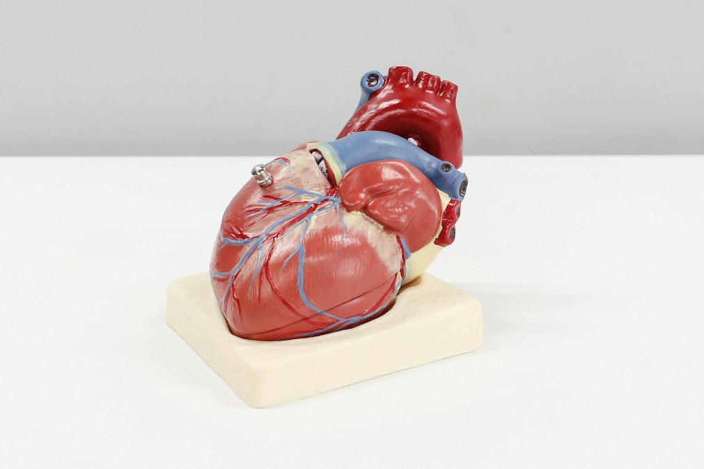
Every heartbeat you have ever had started as an electrical signal, not a mechanical squeeze. That signal originates in a clump of specialized cells the size of a grain of rice, sitting in the upper right chamber of your heart, and it fires roughly 100,000 times a day without any instruction from your brain. Arthur Keith and Martin Flack found this structure in 1907 while dissecting a mole’s heart at University College London, and named it the sinoatrial node. Their discovery answered a question that had puzzled physiologists for decades: what makes a heart, removed from the body entirely, keep beating on its own for a while?
The answer is that the heart is not waiting for permission. It generates and conducts its own electricity, on its own schedule, through a dedicated wiring system built from modified muscle cells that carry current instead of just contracting. Willem Einthoven figured out how to read that electricity from outside the body in 1903, using a string galvanometer in Leiden that weighed as much as a small car, and won the Nobel Prize in 1924 for turning it into the electrocardiogram, the ECG, that every hospital in the world still runs today. What Einthoven’s machine actually records is the heart’s wiring diagram in action, one wave at a time.
Your Heart Runs on a Clock It Built Itself
Here is the part that surprises people: the sinoatrial node does not need your nervous system to tell it to fire. Isolate heart muscle cells in a petri dish, keep them warm and fed, and they will start contracting rhythmically on their own within hours. That is because cardiac cells have an intrinsic electrical instability built into their membranes. Ion channels, mostly ones that let sodium, potassium, and calcium cross the cell membrane, open and close in a predictable sequence that drives the cell’s voltage up, then back down, then up again. In the sinoatrial node specifically, this cycle repeats on its own, generating what physiologists call a pacemaker potential. It is a self-winding clock made of ion channels.
The heart doesn’t wait for the brain’s permission to beat.
Your nervous system still has a say. The vagus nerve slows the sinoatrial node down, which is why a resting heart rate of 60 beats a minute is normal and not a medical emergency. Sympathetic nerves speed it up, which is why your heart rate climbs before you even start running, anticipating the demand. But the vagus and sympathetic inputs are dials, not switches. Cut every nerve connecting the heart to the brain, as happens in a heart transplant, and the transplanted heart still beats. It just cannot speed up in anticipation the way a normally innervated heart does, because it is missing the sympathetic preview signal. That is one of the odd, well-documented quirks of transplant physiology: the new heart works, but it has to actually feel the exercise start before it responds, rather than getting ahead of it.
The Wiring Diagram Nobody Explains to You in Health Class
Once the sinoatrial node fires, the electrical wave has to travel through the rest of the heart in a specific order, or the four chambers stop working together. The signal spreads first across the two upper chambers, the atria, causing them to contract and push blood into the lower chambers. Then it hits a second checkpoint called the atrioventricular node, sitting near the center of the heart, which does something counterintuitive: it slows the signal down on purpose. That delay, on the order of a tenth of a second, gives the atria time to finish emptying before the lower chambers, the ventricles, contract. Skip that delay and the chambers fire almost simultaneously, which is far less efficient at actually moving blood.
From the atrioventricular node, the signal drops into a structure called the bundle of His, named for Wilhelm His Jr., who described it in 1893 while working in Basel. The bundle splits into left and right branches that run down the wall separating the two ventricles, then fan out into a network of fibers named for Jan Evangelista Purkinje, a Czech physiologist who first described them under a microscope in 1839, decades before anyone understood what they actually did. The Purkinje fibers conduct electricity roughly four times faster than ordinary heart muscle, which lets the ventricles contract almost in unison rather than as a slow ripple. The whole trip, from the sinoatrial node’s first spark to the last ventricular fiber, takes well under a second.
A heartbeat is a wave of ions, timed to the millisecond, run through a wiring diagram that took a century to fully map.
What makes this system elegant is that it fails in ways that are almost always locatable. A problem with rate usually traces back to the sinoatrial node. A problem with the handoff between atria and ventricles traces back to the atrioventricular node. A problem with coordinated contraction traces back to the bundle branches or Purkinje network. Cardiologists spend years learning to read an ECG precisely because the tracing tells you not just that something is wrong, but roughly where the wire is broken.
When the Wire Breaks, You Don’t Always Feel It Coming
Arrhythmia is the umbrella term for any disruption to this wiring, and the range is enormous. Some arrhythmias are rate problems: the sinoatrial node fires too slowly, a condition called sinus bradycardia, or a competing group of cells hijacks the rhythm and fires faster than the sinoatrial node can, causing tachycardia. Some are structural: scar tissue from a prior heart attack creates a physical obstacle that electrical signals have to detour around, and that detour can loop back on itself, creating a self-sustaining circuit that fires independently of the sinoatrial node entirely. That looping mechanism, called reentry, is behind a large share of the dangerous ventricular arrhythmias that cause sudden cardiac arrest.
Atrial fibrillation is the most common arrhythmia doctors treat, and it is worth being honest about how much of it remains genuinely unsettled science. The leading model, developed largely from work by Michel Haissaguerre’s group in Bordeaux in the late 1990s, points to rogue electrical signals firing from tissue near where the pulmonary veins enter the left atrium, triggering chaotic, disorganized activity across the rest of the chamber. That model is why catheter ablation, burning or freezing a ring of tissue around those vein openings, has become a standard treatment. But ablation does not cure atrial fibrillation in everyone, success rates depend heavily on how long someone has had the condition, and plenty of cases recur. The underlying question of why some hearts develop these rogue triggers in the first place, and why the disorganized state sometimes sustains itself even after the original trigger is gone, does not have a clean answer yet.
Fixing Broken Wiring With More Wiring
The treatments for electrical heart disease are themselves electrical, which is a fairly direct engineering response to a wiring problem. Wilson Greatbatch built the first practical implantable pacemaker in 1958, in Buffalo, New York, and the story of how he got there is a genuinely famous accident: he reached for the wrong resistor while building a heart rhythm recording device, and the circuit he assembled by mistake produced a pulse pattern that looked exactly like a heartbeat. A modern pacemaker does the same basic job Greatbatch’s device did, generating a small electrical pulse when the heart’s own sinoatrial node fails to fire on time, or when the atrioventricular node fails to pass the signal through. It does not fix the underlying wiring. It substitutes for it.
Michel Mirowski took the idea further at Johns Hopkins, developing the first implantable cardioverter defibrillator, approved by the FDA in 1985, designed to detect the chaotic, disorganized electrical activity of ventricular fibrillation and deliver a shock strong enough to reset every cell in the heart to the same electrical state at once, letting the sinoatrial node take back control. That is the honest, slightly brutal mechanism behind every defibrillator shock: it does not calm the heart down, it stops everything simultaneously so the natural pacemaker gets a clean restart.
Fixing an electrical problem with more electricity isn’t a metaphor here. It’s the actual treatment.
| Year | Development | Who / Where |
|---|---|---|
| 1903 | First practical electrocardiogram | Willem Einthoven, Leiden |
| 1907 | Sinoatrial node discovered | Arthur Keith and Martin Flack, University College London |
| 1958 | First implantable pacemaker | Wilson Greatbatch, Buffalo |
| 1985 | FDA approves implantable cardioverter defibrillator | Michel Mirowski, Johns Hopkins |
| Late 1990s | Pulmonary vein trigger model of atrial fibrillation, basis for catheter ablation | Michel Haissaguerre, Bordeaux |
The Catch Most Pacemaker Patients Never Get Told
A pacemaker fixes timing. It does not fix disease. If your sinoatrial node is failing because of age, or your atrioventricular node is blocked because of scarring from a heart attack, a pacemaker restores the rhythm without touching whatever caused the failure in the first place. That is a real limitation, not a footnote. Patients sometimes hear “pacemaker” and assume their heart problem has been solved in the way a broken bone gets set. What actually happened is that a device is now doing the job a damaged piece of tissue can no longer do reliably, indefinitely, on battery power that eventually needs replacing.
There is a newer wrinkle worth knowing about, too. Traditional pacemakers deliver their pulse to the right ventricle, which is easy to reach but is not where the heart’s own conduction system naturally sends the signal first. Over years, that mismatch can itself weaken the heart’s pumping efficiency, a problem cardiologists call pacing-induced cardiomyopathy. The newer approach, called conduction system pacing, places the lead directly into or near the bundle of His or the left bundle branch, aiming to trigger the heart’s own natural wiring rather than bypass it. It is a smaller, more anatomically precise fix, and it reflects something true about this whole field: the closer treatment gets to mimicking the heart’s original wiring rather than substituting for it, the better the long-term outcomes tend to look.
None of this makes the heart’s electrical system fragile in the way people sometimes assume after a scary ECG reading. It is closer to the opposite: a system stable enough to run for decades on cells that generate their own current, precise enough that a trained eye can localize a fault to a specific junction from a paper tracing, and just imperfect enough that an entire branch of medicine exists to patch the places where the wiring gives out. The open question is not whether we understand the heart’s electricity. It is whether the next generation of devices can talk to that wiring in its own native language, rather than simply overriding it when it fails.
Credit: Ali Hajiluyi on Unsplash