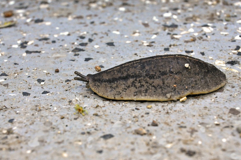
In 2011, in a basement lab at Tufts University in Medford, Massachusetts, a developmental biologist named Michael Levin cut a brown flatworm in half with a scalpel and watched it grow two heads. Nothing about the worm’s DNA had changed. The genome sitting inside every one of its cells still coded for a normal, one headed, one tailed Dugesia japonica. What changed was the worm’s bioelectric state, the pattern of voltage sitting across the membranes of its cells, which Levin’s team had disrupted with a gap junction blocker before the cut was ever made. The worm’s genes were unanimous that it should look like every other planarian on earth. Its electric field disagreed, and the field won.
Planarians have been confusing biologists for more than a century precisely because they refuse to die the way other animals do. Slice one into pieces, and nearly every fragment regrows into a complete worm within about two weeks, each piece producing a head where its front used to be and a tail where its back used to be, no matter where along the body the cut was made. The classic explanation was chemical: gradients of signaling molecules like Wnt proteins, thicker at the tail end and thinner at the head end, supposedly told each fragment which end was which. That explanation is real and it matters. But it cannot be the whole story, because when researchers manipulate voltage alone, without touching Wnt signaling directly, they can override it.
A Voltage Map Sits Underneath the Gene Expression Map
Every animal cell holds a charge across its plasma membrane, the same basic physics that lets a battery do work. Ion channels and pumps push sodium, potassium, chloride, and calcium ions in and out, and the resulting difference in charge, called membrane potential, is usually discussed only in the context of nerve and muscle cells firing electrical impulses. Levin’s lab, along with collaborators including Dany Adams, built a different case over roughly two decades: that this same voltage machinery, present in every cell type, not just excitable ones, forms a slow, stable, body wide map that tells tissue where it is and what it should become.
In planarians specifically, cells at what will become the head end sit at a different resting potential than cells at what will become the tail end, and that difference appears before any visible anatomical change. Block the gap junctions, the protein channels that let adjacent cells share ions and small molecules directly, and the voltage map gets scrambled. Cut the animal while the map is scrambled, and the fragment builds its new head or tail according to the corrupted electrical instructions rather than its original anatomical position.
The genome said one head. The voltage said two. The voltage won.
This is the detail that makes the work more than a curiosity. A gene can specify a protein. It cannot, by itself, specify where in three dimensional space that protein should end up relative to the rest of the body. Something has to tell a mass of genetically identical cells which direction is anterior and which is posterior, and in planarians, that something is measurable in millivolts.
Two Headed Worms That Breed True
The most startling result in Levin’s planarian work was not that two headed worms could be made. Developmental biologists had produced malformed animals through various crude interventions before. The startling part was durability. In a 2011 study published in the International Journal of Developmental Biology, the Levin lab showed that a brief pharmacological disruption of gap junction communication, applied only once, at the time of amputation, produced worms with two heads instead of a head and a tail. Then they cut those two headed worms again, weeks or months later, with no further drug treatment at all. The descendants still grew two heads.
That result does not fit a model where the drug simply damaged tissue or triggered a one time developmental error. Something about the worm’s underlying bioelectric pattern had been reset, not just at the cut site but in a way that persisted through the animal’s ongoing cycle of cellular turnover, and that reset state kept propagating every time the tissue was asked to regenerate again. Levin’s team described the phenomenon as evidence that bioelectric state functions as a kind of stored pattern memory, sitting apart from genetic sequence, capable of overriding default anatomy across multiple rounds of regeneration.
Cut it again months later, with no drug at all. It still grew two heads.
Planarian stem cells, called neoblasts, are the engine that makes any of this possible. They are the only cells in the adult worm’s body capable of dividing, and a sufficiently large fragment of worm carries enough neoblasts to rebuild every tissue type the species has, nerve cords, eyespots, pharynx, gut branches, from scratch. The neoblasts do the physical building. But the voltage pattern appears to be issuing the instructions about orientation, and when those instructions get corrupted, the neoblasts follow the corrupted version faithfully, head for head, eyespot for eyespot, in exactly the wrong place.
Ion Channels, Not Just Signaling Proteins, Set the Pattern
Gap junction blockers are a blunt instrument. They interfere with communication between cells generally, which leaves open the question of whether voltage itself is doing the patterning work or whether the drugs are disrupting something else that happens to travel through the same channels. Later experiments narrowed the case. Researchers targeted specific ion channels and pumps directly, including manipulations of potassium channels and the proton pump V-ATPase, and found that altering ion flow through these specific proteins was enough on its own to shift where regenerating planarian fragments built heads versus tails.
That specificity matters because it rules out a simple alternative explanation, that gap junction blockers were just making cells sick and sickness happened to produce malformed worms. Targeted ion channel manipulation produces targeted, repeatable anatomical outcomes. Push the resting potential of a tissue region in a particular direction, and you can predict, not just observe after the fact, whether that region builds head structures or tail structures. The voltage is not a side effect of the patterning process. It appears to be an instruction set that the patterning process reads.
The same body of research extended well beyond planarians, though the flatworm work remains some of the cleanest demonstration of the principle because the animal regenerates so reliably and so completely. Levin’s lab and collaborators have shown related bioelectric effects shaping craniofacial patterning in frog embryos and limb regeneration in frog tadpoles that normally cannot regrow lost limbs at all. The planarian experiments carry particular force because the comparison is so stark: identical genome, identical cut, two different anatomical outcomes, decided by membrane voltage.
What a Flatworm’s Electric Field Implies About Bigger Animals
None of this suggests that genes are optional. Genes build the ion channels, the pumps, and the gap junction proteins that generate the voltage map in the first place, and without the correct genetic toolkit there would be no bioelectric signal to manipulate. The relationship runs in both directions. Genes build the hardware that produces the voltage pattern, and the voltage pattern then governs how that same genetic hardware gets deployed across the body, which cells differentiate into which tissue type, and where.
The practical interest in this work, for a field like regenerative medicine, is obvious once the mechanism is stated plainly. Humans do not regrow lost limbs or lost organs. Axolotls regrow limbs. Planarians regrow entire bodies from slivers. If anatomical identity in regeneration is substantially a bioelectric decision rather than a purely genetic one, then the lever for inducing regeneration in animals that currently cannot do it might not require rewriting their genome at all. It might require resetting the voltage pattern that genome is already capable of producing, under different circumstances, the way Levin’s tadpole limb work has already begun to suggest.
| Intervention | Mechanism Targeted | Observed Outcome in Planaria |
|---|---|---|
| Gap junction blockade at amputation | Cell to cell electrical and ionic communication | Two headed worms, trait persists through later regenerations without re-treatment |
| Potassium channel modulation | Resting membrane potential of regenerating tissue | Shifted head versus tail identity at the wound site |
| V-ATPase proton pump manipulation | Ion flow and local voltage gradient | Altered anterior-posterior patterning of new tissue |
| No intervention, normal cut | Baseline bioelectric and chemical gradients | Correct head at anterior wound, correct tail at posterior wound |
The Limits of a Flatworm Analogy
It is worth being precise about what the planarian experiments do and do not demonstrate, because the two headed worm photographs tend to travel further than the caveats attached to them. These are worms, not mammals, and planarian neoblasts have regenerative powers that no human stem cell population comes close to matching. Reproducing a bioelectric reset in a mouse limb or a human spinal cord faces an enormous additional problem the flatworm does not: mammalian tissue mounts a scarring response at the wound site that planarian tissue does not, and that scar tissue can physically and electrically block the kind of signaling Levin’s experiments depend on.
Even inside the planarian literature, the precise molecular chain connecting a resting potential difference to the activation of specific head or tail genes is not fully mapped. Researchers know that disrupting voltage changes where Wnt pathway genes and other polarity determining genes get expressed. They do not yet have a complete, step by step account of how an electrical gradient gets translated, inside a cell, into a specific transcriptional decision. The field has strong causal evidence and a plausible general mechanism. It does not yet have the full molecular wiring diagram.
That gap is exactly why the planarian system remains valuable rather than merely historical. It is simple enough, and regenerates reliably enough, to keep serving as the test bed where that wiring diagram gets filled in, one ion channel and one gap junction protein at a time.
A flatworm cut into three pieces does not consult its genome to decide which piece gets a head. It consults a voltage map that existed before the cut, that can be rewritten by a researcher with the right drug, and that will keep producing the rewritten anatomy for generations of cuts to come. The open question is not whether bioelectric fields pattern bodies. Planarians already answered that. The open question is how far up the tree of life that same electrical logic still holds the pen.
Credit: Karl Callwood on Unsplash