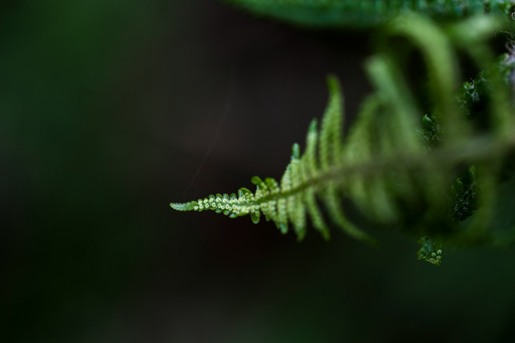
Resurrection fern grows in dense gray-green mats across the branches of live oak trees throughout the American South, curling into a shriveled, seemingly dead husk during drought and unfurling again within hours of rain. Botanists have studied Pleopeltis polypodioides for decades because of that trick, a desiccation tolerance so extreme the plant can lose most of its water content and still recover. That is the organism at the center of a preprint that began circulating among plant biophysicists in the fall of 2024, describing something none of the standard literature on fern physiology predicts: a continuous electrical field detectable in the air and substrate up to 40 centimeters from a single frond.
The claim has not been through peer review. It has not been replicated by an outside lab, at least not yet, and the researchers behind it have been circumspect about the exact recording conditions. But the number itself is what has plant electrophysiologists talking. Forty centimeters is not a subtle signal. It is roughly the distance from a dinner plate to the far edge of a table, measured from a plant organ a few centimeters wide. Nothing in the existing catalog of plant bioelectric behavior reaches that far.
A Field That Does Not Match the Known Physiology
Plants are electrically active. That part is not in dispute and has not been for more than a century. Jagadish Chandra Bose demonstrated in the early 1900s that plant tissue produces measurable electrical responses to touch, injury, and light, work that laid the foundation for what is now called the plant electrome. Alexander Volkov, a biophysicist who has spent much of his career recording action potentials in Venus flytraps and Mimosa pudica, has shown that a flytrap’s trigger hairs generate an action potential of roughly 100 millivolts that propagates across the leaf fast enough to snap the trap shut in under a second. Wounded tomato and Arabidopsis plants send electrical signals through the phloem that trigger defense gene expression in leaves the injury never touched.
What none of that work describes is a field that projects outward from the plant into open space or soil at a stable amplitude across tens of centimeters. Plant electrical signals are typically intracellular or travel cell to cell through plasmodesmata and vascular tissue. They decay quickly outside the organism because the signal depends on ion channels and membrane potentials inside living cells, not on a broadcast field the way a neuron’s synapse can trigger activity across a gap. A field with the reach described in the fern preprint would need a different explanation altogether, and the preprint offers only a partial one.
Forty centimeters is not a subtle signal. Nothing in the existing literature on fern physiology reaches that far.
What the Preprint Actually Measured
The recording setup described in the document uses a pair of non-invasive surface electrodes placed at increasing distances from a fern frond, with a reference electrode in the surrounding soil. The reported field is described as sustained rather than transient, meaning it did not spike and decay the way an action potential does, but persisted at a measurable amplitude for the duration of the recording sessions, which the authors describe in hours rather than minutes. The preprint does not specify the field strength at the 40 centimeter mark, and that omission matters. A field of a few microvolts at that range is a very different claim from one measured in millivolts, and the difference changes what kind of biological or non-biological process could plausibly generate it.
Andrew Adamatzky, a computer scientist at the University of the West of England who has published extensively on electrical spiking in fungal mycelium networks, has argued in his own work that living systems produce far more electrical complexity than biology has historically bothered to measure. His 2022 paper on fungal electrical activity described spiking patterns in mycelium that resembled, at a crude level, patterns of neural firing, though he was careful to note the comparison was structural, not functional. The fern claim sits in the same territory of plausibility that Adamatzky’s fungal work occupies: an unusual electrical signature in a non-neural organism that invites comparison to nervous systems it almost certainly does not have.
The Confounds a Field This Large Would Need to Rule Out
Extracellular electrical measurements on living tissue are notoriously easy to contaminate. Soil moisture gradients generate streaming potentials on their own, produced by water moving through porous material and dragging charge with it, entirely independent of anything the plant is doing. Humidity differences near a transpiring frond can create capacitive coupling between electrodes that mimics a biological signal. Electrode drift, a slow baseline shift caused by chemical reactions at the metal contact surface, can produce a reading that looks sustained over hours when it is really just the electrode degrading at a steady rate.
None of this means the fern finding is wrong. It means the finding, as currently described, has not eliminated the alternatives that would need to be eliminated before a plant physiologist accepts that Pleopeltis polypodioides is generating a genuine 40 centimeter bioelectric field rather than an artifact of how the measurement was taken. A properly controlled version of this experiment would run in a Faraday cage, use electrodes calibrated against a dead or excised frond as a negative control, and vary soil moisture independently of the fern’s physiological state to see whether the field tracks the plant or tracks the substrate.
The finding has not eliminated the alternatives it would need to eliminate to be believed.
Why Bioelectricity Researchers Are Paying Attention Anyway
Even skeptical readers of the preprint are not dismissing it outright, and the reason has to do with a broader shift in how biologists think about bioelectric signaling generally. Michael Levin’s lab at Tufts has spent the past decade demonstrating that bioelectric gradients across non-neural tissue, not just neurons, direct pattern formation in developing embryos and regenerating limbs. That work reframed voltage as a signal available to essentially any tissue with a membrane, not a specialized trick reserved for nerve and muscle. If bioelectric fields do more organizing work across the tree of life than biology assumed a generation ago, a fern producing an unexpectedly extended field is not automatically absurd. It is just unverified.
There is also a useful contrast with organisms that unambiguously do generate large external electrical fields for a functional reason. Electric fish in the genus Gymnotus produce weak electric fields around their bodies for electrolocation, sensing objects and other fish through distortions in a self-generated field that typically extends tens of centimeters, in the same range claimed for the fern. The electric eel, Electrophorus electricus, generates discharges up to roughly 600 volts for predation and defense using dedicated electrocyte tissue evolved specifically for that purpose. Ferns have nothing resembling electrocytes. If the 40 centimeter field is real, it would not be doing the same job electrolocation does in a weakly electric fish, because a fern has no nervous system to interpret a return signal. What it would be doing is an open question the preprint does not answer.
| Organism or system | Typical signal | Approximate range |
|---|---|---|
| Venus flytrap action potential | ~100 mV, transient | Within the leaf |
| Human surface ECG | ~1 mV at skin | Local, chest surface |
| Gymnotus electric fish (electrolocation) | Weak field, continuous | Tens of centimeters |
| Electric eel discharge | Up to ~600 V, pulsed | Meter scale, in water |
| Resurrection fern (preprint claim) | Amplitude not disclosed, sustained | Up to 40 cm (unreplicated) |
What Verification Actually Looks Like From Here
Bioelectricity as a field has been burned before by findings that generated headlines faster than they generated replication. The path from a striking preprint to accepted science runs through independent labs reproducing the measurement under conditions the original team did not design, ideally without knowing what result they are supposed to find. For a claim this specific, that means at minimum one lab outside the original group running the Faraday cage and dead-tissue control experiments described above, disclosing raw field strength data rather than a distance threshold alone, and testing whether the effect appears in other fern species or is unique to Pleopeltis polypodioides and its unusual desiccation biology.
None of that has happened yet. What has happened is that a preprint describing an anomalous electrical measurement in a common Southern fern has reached researchers who study bioelectric signaling in flytraps, fungi, and embryos, and none of them are willing to call it impossible. That is a lower bar than confirmation, but it is not nothing. The question the fern raises is not whether plants can be electrically interesting, decades of work already answered that. The question is whether this particular electrical signature belongs to the fern at all, or to the soil, the humidity, and the electrodes sitting in it.
Credit: Diana Parkhouse on Unsplash