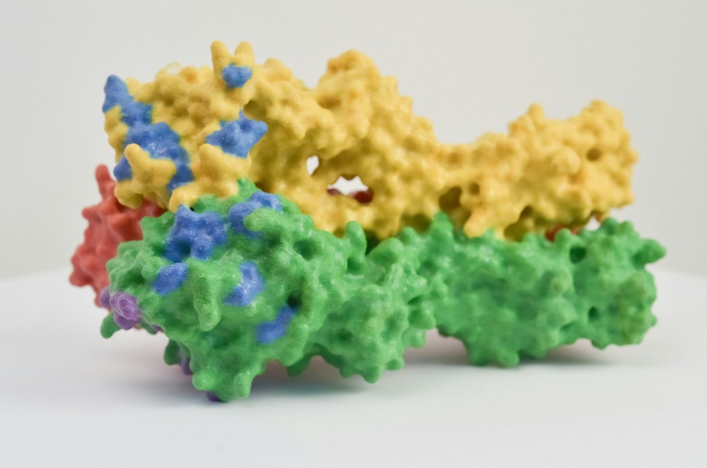
In the spring of 1998, a structural biologist named Roderick MacKinnon walked his team at Rockefeller University through a set of X-ray diffraction images that would earn him the Nobel Prize in Chemistry five years later. The images showed, for the first time at atomic resolution, the shape of a potassium channel called KcsA, borrowed from a soil bacterium called Streptomyces lividans. It looked like a tiny inverted teepee, four identical protein subunits arranged around a central pore barely wide enough for a single potassium ion to pass through single file. At the narrowest point sat a ring of oxygen atoms, spaced with almost exact geometric precision, positioned to mimic the water molecules that normally surround a potassium ion in solution.
That ring solved a problem that had puzzled physiologists for four decades. Potassium channels let potassium ions through at a staggering rate, close to the physical limit of diffusion, while blocking sodium ions almost completely, even though sodium is smaller. MacKinnon’s structure showed why size alone doesn’t decide the outcome. The channel doesn’t act like a screen door sorting objects by width. It acts like a chaperone, stripping water molecules off an approaching ion and replacing them with an equivalent cage of oxygen atoms, but only if the ion is exactly the right size for that substitution to be energetically free. Sodium is too small. The oxygen ring can’t hug it closely enough, and the energy cost of dehydrating it never gets paid back. Potassium fits perfectly, so it slides through nearly a thousand times faster.
That single structure, from a bacterium, explained a property found in every nerve cell in the human brain, and it captured what an ion channel actually is: not a hole in a membrane, but a molecular machine that reads its cargo before deciding whether to let it pass.
A Pore That Reads Its Guest List
Every cell in the body is wrapped in a lipid membrane, a double layer of fat molecules that is, on its own, essentially impermeable to charged particles. Sodium, potassium, calcium, and chloride ions cannot cross a naked lipid bilayer at any biologically useful rate. That impermeability is what makes a membrane useful for storing electrical charge in the first place, the same way a plastic-coated wire holds current instead of leaking it. But a membrane that stores charge perfectly is useless for signaling unless something can open a controlled path across it at a chosen moment. That something is the ion channel: a protein, or more often a complex of several proteins, embedded across the membrane with a water-filled pore running through its center.
The pore reads its guests before it lets them in.
Ion channels are named for what they let through. Sodium channels, potassium channels, calcium channels, and chloride channels each have their own protein family, their own gene lineage, and their own selectivity filter shaped to favor one ion species over the others by a factor of hundreds or thousands. Bertil Hille, whose work at the University of Washington helped define much of modern channel physiology starting in the 1970s, described this as one of the central design principles of excitable membranes: separate channels for separate ions, each independently switchable, so a cell can shape a precise electrical event instead of just leaking charge indiscriminately.
Voltage Is a Sensor, Not Just a Consequence
Selectivity explains what gets through. It doesn’t explain when. The answer to that question came out of Plymouth, England, in 1952, when Alan Hodgkin and Andrew Huxley published a series of papers describing electrical recordings from the giant axon of a squid, a nerve fiber wide enough, nearly a millimeter across, to thread with a wire electrode using the equipment available at the time. Hodgkin and Huxley never saw a channel protein; those wouldn’t be identified for another three decades. But their equations, derived purely from measured current and voltage, predicted that the axon’s membrane contained two distinct populations of gates: one that opened quickly in response to voltage and let sodium rush in, and another that opened more slowly and let potassium rush out. Their model reproduced the action potential almost perfectly, and it won them the Nobel Prize in 1963.
What Hodgkin and Huxley called gates, later biophysicists identified as a physical structure inside the channel protein itself: the voltage sensor, a cluster of positively charged amino acids arranged on a helix that sits within the membrane’s electric field. When the membrane depolarizes, that field shifts, and the charged helix physically moves, dragging the rest of the protein into a new shape and swinging the pore open. Clay Armstrong’s work in the 1970s, measuring the tiny “gating currents” produced by that charge movement before the ion current itself began to flow, gave the first direct experimental evidence that the sensor was a real, moving piece of protein rather than a mathematical convenience.
A channel doesn’t just open. It decides, in a fraction of a millisecond, what is allowed through.
This is the mechanism that makes voltage-gated sodium channels the trigger for every action potential in the nervous system. A stimulus depolarizes the membrane a few millivolts. Sodium channels sense the change and snap open within microseconds, sodium ions flood in, and the membrane depolarizes further still, snapping open more channels in a self-reinforcing cascade. Within a millisecond the same channels inactivate, a separate part of the protein swinging in to plug the pore from the inside, cutting the sodium current off before the cell can overshoot. Potassium channels open slightly later and stay open longer, pulling the membrane back down to rest. The whole cycle, from trigger to recovery, takes one to two milliseconds. It is the electrical unit that every thought, reflex, and heartbeat is built from.
One Channel, One Ion, One Job
The human genome encodes more than 140 distinct voltage-gated ion channel genes, plus a separate large family of ligand-gated channels that open not in response to voltage but to a chemical messenger binding directly to the protein: acetylcholine at the neuromuscular junction, glutamate and GABA at synapses throughout the brain. Each gene product is tuned for a specific job. Cardiac sodium channels (encoded by SCN5A) activate and inactivate on a different timescale than the brain’s sodium channels (SCN1A, among others), which is part of why a drug can be designed to calm an overactive heart rhythm without silencing the nervous system, or vice versa. Local anesthetics like lidocaine work by wedging into the pore of voltage-gated sodium channels from the inside of the cell, physically blocking the channel in its open or inactivated state and preventing the nerve from firing at all, which is the entire mechanism behind numbing a tooth or a surgical site.
That specificity, gene by gene, tissue by tissue, is what makes ion channels such an attractive and such a difficult drug target. A compound that blocks one calcium channel subtype to treat chronic pain needs to leave the nearly identical calcium channel subtype that keeps the heart beating completely alone. Getting that selectivity right, at the level of a single amino acid difference in a pore, is most of what makes channel-targeted drug discovery slow.
Listening to a Single Molecule
For most of the twentieth century, physiologists could only measure the combined electrical behavior of thousands of channels firing together across a whole cell. That changed in 1976, when Erwin Neher and Bert Sakmann, working at the Max Planck Institute for Biophysical Chemistry in Göttingen, developed the patch clamp technique: pressing a fire-polished glass pipette against a cell membrane, forming a seal tight enough, in the billions of ohms of resistance, to isolate a patch of membrane containing perhaps one or two channel proteins. For the first time, researchers could watch a single ion channel open and close in real time, recording currents measured in picoamps, trillionths of an amp, as individual channels flickered between open and closed states over milliseconds. Neher and Sakmann won the Nobel Prize in Physiology or Medicine in 1991 for the technique, which remains the standard method for characterizing channel behavior in labs studying everything from epilepsy to cardiac arrhythmia to the pharmacology of new anesthetic drugs.
What that single-channel view revealed was more surprising than a smooth, graded response. Individual channels don’t partially open. They flip, stochastically, between fully open and fully closed, staying in each state for a random interval governed by probability rather than a fixed timer. The smooth current recorded from a whole cell is really an average over thousands of these binary, noisy, individual decisions, the same way a smooth tide is really billions of individual water molecules moving independently.
When the Gate Breaks
Because ion channels are single gene products with precisely defined jobs, a mutation in one channel gene tends to produce a specific, often severe, and often well characterized disease. These are called channelopathies, and they span cardiology, neurology, and beyond.
| Channel / Gene | Ion | Associated Condition |
|---|---|---|
| KCNQ1 / KCNH2 | Potassium | Long QT syndrome, cardiac arrhythmia |
| SCN1A | Sodium | Dravet syndrome, childhood epilepsy |
| SCN5A | Sodium | Brugada syndrome |
| CFTR | Chloride | Cystic fibrosis |
| CACNA1A | Calcium | Familial hemiplegic migraine, episodic ataxia |
CFTR is a useful edge case, because it shows the concept stretching past the textbook definition. It is technically a chloride channel, but it is regulated by ATP hydrolysis rather than voltage or a fast-binding ligand, and its malfunction in cystic fibrosis doesn’t produce an electrical symptom so much as a mechanical one: without properly regulated chloride and water transport across epithelial cells lining the lungs and pancreas, mucus thickens and clogs the airways. The drug Trikafta, approved by the FDA in 2019, works by directly correcting the folding and gating defects in mutant CFTR protein, an ion channel drug in the most literal sense, not one that blocks a channel but one that repairs it.
Long QT syndrome and Brugada syndrome sit closer to the classic electrical story. Both involve mutations that throw off the precise timing of sodium or potassium currents during a single heartbeat, either delaying repolarization or accelerating it, and both can produce a dangerous and sometimes fatal arrhythmia in patients who otherwise appear perfectly healthy. Cardiologists screen for these mutations specifically because the underlying defect is not in the heart’s structure. It’s in a single protein’s timing.
The Bridge to Bioelectronic Medicine
Every device covered under the banner of bioelectronic medicine, from a vagus nerve stimulator to a cochlear implant to an experimental brain-computer interface, ultimately works by manipulating ion channels, either directly with an electric field or indirectly by triggering the release of a neurotransmitter that opens a ligand-gated channel downstream. A stimulating electrode doesn’t inject thought or sensation into tissue. It changes the local voltage enough to nudge voltage-gated sodium channels open, triggering the same all-or-nothing action potential that Hodgkin and Huxley described in a squid axon in 1952. The electrode is new. The mechanism it exploits is not.
That continuity is worth sitting with. The gap between MacKinnon’s 1998 crystal structure and a 2024 spinal cord stimulator sold by a medical device company is enormous in engineering terms, and close to nothing in mechanistic terms. Both depend on the same oxygen ring, the same charged helix, the same millisecond cascade of opening and closing that a cell membrane has been running for as long as there have been cells with membranes.
An ion channel, in the end, is a small enough thing to hold at atomic resolution in a single crystallography image, and a large enough idea to explain a heartbeat, a memory, a local anesthetic, and a genetic disease with the same handful of physical rules. Every field that claims to work with bioelectricity, medicine included, is really just finding new ways to ask that same molecular gate to open.
Credit: National Institute of Allergy and Infectious Diseases on Unsplash