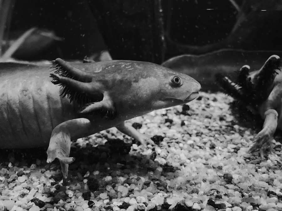
Cut off an axolotl’s leg and the stump starts generating its own electricity before it starts growing new tissue. Not metaphorically. A measurable current, flowing out of the wound, detectable with a vibrating probe electrode held a few micrometers from the skin. That current shows up within minutes of amputation and persists for weeks, long before anyone can see a blastema forming under the microscope. I keep coming back to this fact because it inverts the story most people have in their heads about regeneration, which is that it is fundamentally a genetic program. It is not, or at least not only. The animal is running a physics experiment on itself before it runs a genetics one.
The technique that first caught this current, the vibrating probe, came out of work by Lionel Jaffe and Richard Nuccitelli in the 1970s, and it let researchers measure extracellular electric fields around living cells without touching them. Richard Borgens, working at Purdue University, used it on regenerating newt and axolotl limb stumps and found currents on the order of tens of microamps per square centimeter flowing outward through the wound epithelium, a layer of skin cells that seals the stump within hours of injury. That layer is not passive scar tissue. It is an electrically active membrane, pumping sodium ions inward and generating a steady voltage difference between the stump and the surrounding intact skin. Borgens called it a skin battery, and the name has stuck because it is literally accurate.
The Stump Behaves Like a Battery Before It Behaves Like a Program
What made the injury current interesting to bioelectricity researchers, rather than a curiosity, was the question of causation. Correlation between electric current and regeneration outcome is easy to observe and easy to dismiss. Borgens, along with John Vanable and Jaffe, ran the experiment that mattered: block the current pharmacologically, without touching a single gene, and see what happens to the limb. They applied amiloride, a drug that blocks epithelial sodium channels, directly to regenerating amphibian stumps. Sodium influx through those channels is what generates the outward current in the first place. With amiloride on board, the current collapsed, and blastema formation, the mass of dedifferentiated cells that would otherwise rebuild the limb, failed to organize properly.
The stump runs a physics experiment before it runs a genetics one.
That result is the load-bearing evidence for everything that follows in this field. It demonstrates necessity, not just association. A regenerating limb does not merely produce electric current as a side effect of wound healing. It requires that current, or something the current does, to complete the regenerative sequence. And critically, none of this involved editing a genome, inserting a transgene, or knocking out a gene with CRISPR. It involved a small-molecule ion channel blocker applied topically to intact tissue in an intact animal. The intervention was electrochemical, not genetic, and the effect was still developmental.
What the Voltage Gradient Actually Encodes
The mechanism runs through resting membrane potential, the voltage difference across a cell’s outer membrane that most biology courses mention once and then move past. In a regenerating limb, cells in and around the wound do not sit at a single uniform voltage. They form a gradient, with more depolarized cells near the amputation plane and more polarized cells further into intact tissue. That gradient is set by the distribution and activity of ion channels and pumps, sodium and potassium channels, the H+ V-ATPase proton pump, and gap junctions, the direct channels that let adjacent cells share ions and small molecules and effectively vote on their collective voltage state.
Michael Levin, who runs the Allen Discovery Center for Regenerative and Developmental Biology at Tufts University, has spent two decades building the argument that these voltage gradients function as a kind of pre-pattern, a physical layout of positional information that tells cells what structure they are supposed to become before the relevant genes are transcribed. His group has shown in frog and planarian systems that shifting bioelectric state with ionophores and channel-targeted drugs can redirect what regenerates, producing outcomes like ectopic eyes or two-headed flatworms without introducing new genetic material. The axolotl work sits downstream of that framework. The animal already runs the program Levin’s lab is trying to describe. The question for axolotl researchers has been whether the same categories of pharmacological tools, sodium channel blockers, gap junction inhibitors, ion pump modulators, can reveal and redirect the same kind of gradient in a system that actually regenerates a full limb on its own, rather than just a tail or a flatworm fragment.
Specificity in regeneration comes from voltage, not just from which genes turn on.
Gap junction blockers make the point cleanly. Compounds like heptanol and carbenoxolone sever the electrical coupling between blastema cells without altering their DNA. Disrupt that coupling during the critical early window after amputation, and the blastema loses coordination. Cells proliferate, but they do not organize along a consistent proximal-distal axis, the line running from shoulder to fingertip that determines whether the animal regrows a full limb or a disorganized mass of cartilage and skin. The voltage gradient, in other words, does not just accompany pattern formation. It appears to specify it.
Why Pharmacology Beats Gene Editing Here
Axolotls have resisted genetic manipulation far longer than the standard laboratory animals of developmental biology. Their genome, sequenced by Sergej Nowoshilow, Elly Tanaka, and colleagues and published in Nature in 2018, runs to roughly 32 billion base pairs, about ten times the size of the human genome, bloated with repetitive sequence and long introns that make CRISPR-based knockouts slower and messier to validate than in mice or zebrafish. That practical obstacle has quietly pushed axolotl regeneration research toward physiological interventions, not out of any grand strategic preference for bioelectricity as a discipline, but because dosing a stump with amiloride or ouabain is achievable in a week, while generating and screening a stable axolotl knockout line can take a year or more given the animal’s slow generation time and enormous genome.
The side effect of that constraint has been methodologically useful. Pharmacological and biophysical tools, topical drugs, externally applied electric fields, ion-selective microelectrodes, work on wild-type animals in real time, and they are reversible in a way gene editing is not. Reverse an externally applied field around a regenerating newt or axolotl limb, an experiment with roots going back to work by Gerald Marsh and Harold Beams in the mid-twentieth century on directing regeneration polarity with applied current, and you can watch the tissue’s growth axis respond within days. Wash out the drug, and channel activity returns to baseline. That kind of clean, temporary, dose-dependent control over a developmental outcome is difficult to achieve with a permanent genetic edit, and it is part of why this line of research has stayed relevant even as gene-editing tools have gotten easier to use elsewhere in biology.
| Intervention | Mechanism | Effect on regeneration |
|---|---|---|
| Amiloride | Blocks epithelial sodium channels | Collapses stump current, impairs blastema formation |
| Ouabain | Inhibits Na/K-ATPase pump | Flattens transmembrane voltage gradient, halts regeneration |
| Heptanol / carbenoxolone | Blocks gap junction coupling | Disorganized blastema, loss of proximal-distal patterning |
| Externally applied field reversal | Overrides endogenous current direction | Redirects regeneration axis and nerve sprouting orientation |
The Comparison to Mammals Is the Real Point
Humans do not lack these ion channels. We have sodium channels, potassium channels, gap junctions, the same basic electrochemical hardware axolotls use to rebuild a leg. What we lack is the wound epithelium behavior that keeps the current flowing. A mammalian wound closes by forming a keratinized scab and eventually scar tissue, an electrically resistive barrier that shuts the current down within days. An axolotl wound epithelium stays thin, ion-permeable, and electrically active for weeks, long enough for the voltage gradient to do its patterning work underneath. That difference in wound epithelium physiology, not some absent regeneration gene, is one of the leading candidate explanations for why a newt regrows a limb and a person grows a scar.
Randal Voss, who runs the Ambystoma Genetic Stock Center at the University of Kentucky and supplies much of the world’s research colony of axolotls, has built the genetic and genomic infrastructure that lets labs cross-reference bioelectric findings against gene expression data at unprecedented resolution for this species. That resource matters because the two approaches, bioelectric and genetic, are not competitors. The voltage gradient appears to act upstream of transcriptional changes, altering which genes turn on by altering the physical and ionic environment those genes are read in. Mapping where the two causal chains meet, exactly which channels create the gradient and exactly which genes respond to it, is the unfinished part of this story.
Where the Evidence Still Runs Thin
I want to be direct about the limits here rather than round them off. Most of the causal, drug-based evidence for bioelectric control of regeneration comes from newt limbs and Xenopus tadpole tails rather than axolotl limbs specifically, and axolotl-specific bioelectric mapping, tracking the precise voltage gradient across a regenerating axolotl blastema in real time with modern voltage-sensitive dyes, is thinner in the published literature than the broader theoretical framework would suggest it should be. The field has good reason to expect the same logic applies across salamander species, since the injury current and wound epithelium physiology are broadly conserved among them, but “should apply” and “has been directly measured” are different claims, and this is a place where the axolotl-specific data still needs filling in rather than assumed by analogy.
That gap is itself the interesting research frontier right now, more interesting to me than another genetic knockout screen would be. If the voltage gradient really does sit upstream of gene expression in axolotl limb regeneration the way it does in frog tail regeneration, then the practical path toward provoking regeneration in animals that do not naturally do it, mice, eventually humans, runs through pharmacology and bioelectric engineering rather than gene therapy. You cannot gene-edit a human patient’s entire limb stump. You can, in principle, apply a topical ion channel drug or an external field to one. Nobody has done that successfully in a mammal yet. But the axolotl has already shown, without a single genetic modification, that the electricity comes first.
Credit: Tommy Bond on Unsplash