Researchers working on bladder dysfunction after spinal cord injury have gotten a closed-loop nerve implant to do something sacral neuromodulation devices on the market today cannot: restore the timed, coordinated signaling between the bladder muscle and the urethral sphincter in rats, not just suppress the overactivity that makes neurogenic bladder so miserable to live with. The work sits in the research tradition built by labs at Duke University, the University of Michigan, and Case Western Reserve University, all of which have spent years mapping how the sacral spinal cord and the pudendal nerve talk to the bladder, and what happens to that conversation when a spinal injury cuts the line to the brain.
The headline result is narrow and rodent-sized. But the mechanism behind it is the part worth your attention, because it points at a different philosophy for treating bladder dysfunction than the one behind every FDA-approved device currently implanted in patients.
The Injury Doesn’t Break the Bladder, It Breaks the Timing
After a spinal cord injury above the sacral segments, the bladder and the sphincter muscle around the urethra don’t stop working. They stop talking to each other. Normally, the brain holds the bladder’s emptying reflex in check until you decide otherwise, then releases it in a coordinated sequence: the detrusor muscle contracts while the sphincter relaxes, so urine actually leaves the body instead of getting forced against a closed valve. Cut the spinal cord above the sacral cord, and that top-down coordination disappears. The local sacral reflex circuit still fires, but now it fires on its own schedule, and often the detrusor contracts while the sphincter stays clenched, a condition called detrusor-sphincter dyssynergia.
The injury doesn’t destroy the bladder circuit. It cuts the brain out of the conversation.
That mismatch is not a minor inconvenience. High bladder pressure with no outlet pushes urine backward toward the kidneys, and over years that backflow is a leading cause of kidney damage in people living with spinal cord injury. It also triggers autonomic dysreflexia, a sudden, dangerous spike in blood pressure that can be triggered by something as routine as a full bladder. Intermittent self-catheterization, done several times a day, remains the standard way to manage this. It works, but it is invasive, it carries a steady risk of urinary tract infection, and it requires either the patient or a caregiver to do it on a clock, every day, for life.
Why Sacral Neuromodulation Isn’t the Same Thing as Fixing the Circuit
The device most people have actually heard of here is Medtronic’s InterStim, a sacral nerve stimulator originally approved by the FDA in 1997 for urinary urge incontinence and later expanded to urinary retention. Axonics Modulation Technologies brought a rechargeable competitor to market in 2019. Both work by placing a lead near the S3 sacral nerve root and delivering continuous or scheduled electrical pulses to calm an overactive bladder reflex.
These devices are real medicine, used in hundreds of thousands of patients, and they work well for a specific population: people whose bladder is overactive but whose basic wiring between detrusor and sphincter is otherwise intact. They are much less effective for the dyssynergia problem that follows spinal cord injury, because they are not actually coordinating two muscle groups. They are turning a dial down on a nerve and hoping the whole system calms along with it.
Sacral neuromodulation dampens the signal. It doesn’t restore the dialogue between detrusor and sphincter.
Warren Grill’s lab at Duke has spent much of the last two decades on a more targeted idea: use electrical stimulation not as a blunt suppressor but as a substitute for the missing supraspinal signal, timed specifically to make the sphincter relax at the moment the bladder contracts. Tim Bruns’s group at the University of Michigan has pushed a related approach, recording directly from the pudendal nerve to sense bladder filling in real time, then using that signal to decide when and how to stimulate, rather than stimulating on a fixed schedule regardless of what the bladder is actually doing. Narendra Bhadra and Kevin Kilgore’s work at Case Western Reserve added another piece: high-frequency alternating current can be used to temporarily block nerve conduction, not just excite it, which means a single cuff electrode can, in principle, both trigger a contraction and hold a sphincter open at the same time, something a simple stimulating electrode cannot do on its own.
What the Cuff Electrode Actually Has to Do
The newer closed-loop work merges these three ideas into one device: a multi-contact nerve cuff placed on sacral spinal nerve roots or the pudendal nerve, wired to electronics that read afferent signals correlating with bladder pressure and decide, moment to moment, what pattern of stimulation to send back. Instead of a stimulator cycling on a timer, you get something closer to a thermostat: sense the state of the system, then apply exactly the correction needed, and nothing more.
In rats with a spinal injury, that meant the implant could pick up on rising bladder pressure as the organ filled, hold off until pressure crossed the threshold associated with the voiding reflex, then deliver a stimulation pattern tuned to drive detrusor contraction while suppressing sphincter activity long enough for the bladder to actually empty. That is a fundamentally different task than what InterStim and Axonics do. It is also a much harder engineering problem, because it requires the implant to both listen and speak on the same nerve, in real time, without the two functions interfering with each other.
This is where you should pay attention to the catch, because there is a real one. Everything described above happened in rats, most likely under controlled, relatively short-duration testing conditions rather than months of free, unsupervised living. Rat pudendal and sacral nerves are a fraction of a millimeter in diameter, carrying far fewer fibers than the equivalent human nerves, and a cuff electrode that sits cleanly and selectively on a rat nerve does not automatically scale up to a human one. Human peripheral nerves are thicker, more heterogeneous, and surrounded by more connective tissue, all of which make it harder to selectively activate just the fiber population you want without recruiting neighbors you don’t.
The Scar Tissue Problem Nobody Markets
Every chronic nerve implant faces the same slow enemy: the body’s own healing response. Scar tissue, known in this context as fibrotic encapsulation, builds up around an implanted electrode over months, gradually increasing the impedance between the electrode contact and the nerve fibers it’s supposed to read or stimulate. Signals that were crisp in week one get noisier by month six. This is a known limitation in essentially every chronic peripheral nerve interface, from cochlear implants to the Utah array used in brain-computer interface research, and bladder control implants are not exempt from it.
For a device whose entire value proposition is precise, closed-loop timing between two muscle groups, signal degradation is not a cosmetic problem, it is the problem. A sacral neuromodulator that drifts out of calibration over years still mostly works, because its job is relatively coarse: calm the nerve down. A closed-loop dyssynergia implant that drifts out of calibration risks recreating the exact mistimed contraction it was built to fix, possibly without the clinician or patient immediately realizing the device has degraded rather than simply stopped helping.
Where This Sits Against What’s Already Implanted in People
| Approach | Mechanism | Status | Main Limitation |
|---|---|---|---|
| Intermittent self-catheterization | Mechanical drainage, no nerve involvement | Standard of care | Invasive, infection risk, daily burden |
| Medtronic InterStim | Continuous sacral nerve (S3) stimulation | FDA approved, 1997 | Dampens reflex, doesn’t coordinate muscles |
| Axonics system | Rechargeable sacral neuromodulation | FDA approved, 2019 | Same underlying limitation as InterStim |
| High-frequency pudendal block (Case Western research) | Nerve conduction block to relax sphincter | Preclinical / early human studies | Blocks, doesn’t independently drive contraction |
| Closed-loop sacral/pudendal implant | Senses bladder state, times stimulation to coordinate both muscles | Rat studies | Unproven at human nerve scale and over years |
What Would Actually Have to Happen Next
The honest path from this result to a patient’s spine runs through several years of unglamorous engineering work: electrodes that can selectively address specific fiber populations in a human-sized nerve, implant electronics that can run a sensing and stimulating loop for years without drifting, and power systems small enough to sit near the sacrum without becoming their own surgical complication. None of that is exotic by the standards of bioelectronic medicine. Cochlear implants and deep brain stimulators have solved versions of the longevity problem already. But bladder control adds a wrinkle those devices don’t have, because the signal you’re trying to read and the signal you’re trying to send are both happening on the same nerve, in the same window of time, and the quality of the coordination is the entire therapeutic value.
A rat’s pudendal nerve is not a rehearsal for a human one, just a smaller model of the same unsolved problem.
What makes this line of research worth tracking isn’t that it beats catheterization today. It doesn’t, not yet, not in a rat. It’s that it reframes neurogenic bladder as a signal-timing problem rather than an overactivity problem, which is a more accurate description of what spinal cord injury actually does to the circuit. The field has spent two decades building devices that turn the bladder’s reflex down. The open question now is whether anyone can build one that teaches the detrusor and the sphincter to talk to each other again, on a schedule the brain used to set and can no longer reach.
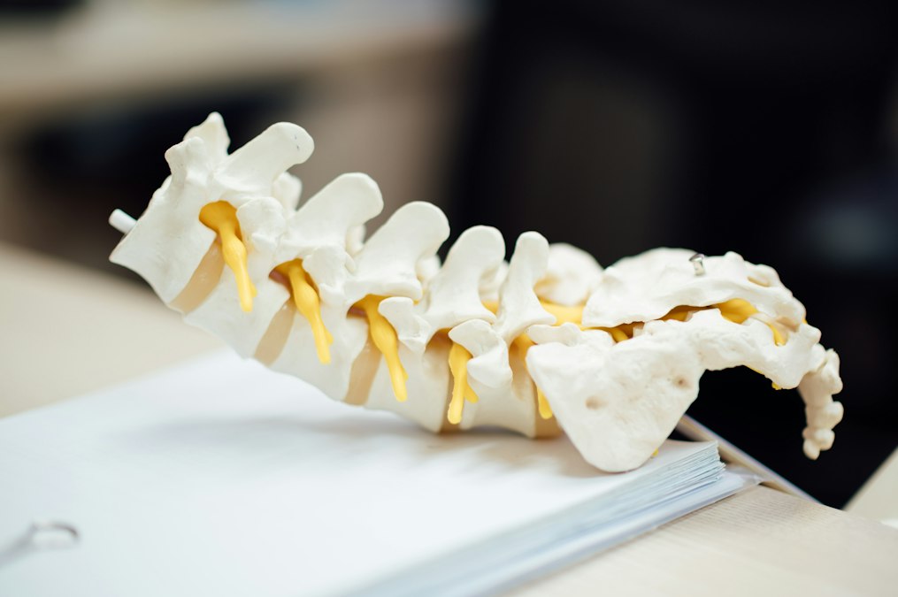
Credit: CHUTTERSNAP on Unsplash