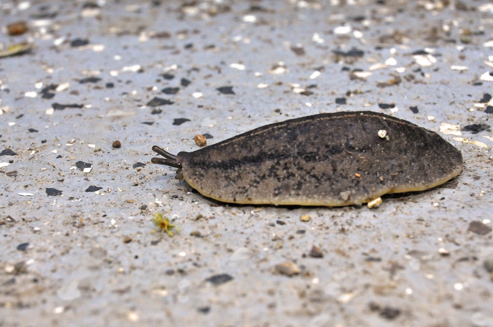
In 2013, in a basement lab at Tufts University, a researcher named Tal Shomrat ran a small flatworm through a training course, cut off its head, and waited two weeks to see if it still remembered anything. The worm was a brown planarian, Dugesia japonica, a species that has been a staple of regeneration biology since the late nineteenth century. Shomrat and his advisor, developmental biologist Michael Levin, had trained groups of these worms to associate a textured, illuminated environment with food. Then they decapitated them. Planaria regenerate a complete new head, including a new brain, from a severed body fragment in about a week to ten days. The question was whether the worm that emerged from that regrowth would behave like it had learned anything at all.
It did. Worms that had been trained before losing their heads reached food faster after regeneration than worms trained for the first time. The original brain, the tissue that presumably held the memory, was gone. A new one had grown in its place from undifferentiated stem cells called neoblasts. Yet something about the prior experience persisted through that reconstruction. The result, published in The Journal of Experimental Biology under the title “An automated training paradigm reveals long-term memory in planaria and its persistence through head regeneration,” did not fit comfortably into a century of memory research built almost entirely around synapses.
A Flatworm That Rebuilds Its Own Brain
Planaria have been useful to biologists precisely because they cheat death in ways vertebrates cannot. Thomas Hunt Morgan documented their regenerative capacity as far back as 1898, cutting worms into dozens of pieces and watching each fragment rebuild a complete animal, head, tail, and all. The organism’s body is packed with neoblasts, adult pluripotent stem cells that can become any cell type the worm needs. Slice a planarian in half and the tail piece grows a new head within roughly a week. Slice it into a dozen pieces and, within limits, each piece can do the same thing.
That regenerative machinery makes planaria a strange kind of natural experiment for memory research. In mammals, removing the brain removes the memory, full stop, because there is no biological process that rebuilds a hippocampus from spare parts. In planaria, the brain is not a fixed structure. It is more like a renewable one, assembled fresh from stem cells according to instructions the worm’s body already carries. Levin’s lab, which now runs the Allen Discovery Center at Tufts, had spent years before the 2013 study establishing that those instructions are not purely genetic. They are, in large part, bioelectric.
Training a Worm, Then Removing Its Head
The training paradigm itself was simple by design. Worms were placed in a petri dish with a textured floor and exposed to a combination of light and a food stimulus at a specific location, conditions that untrained planaria find mildly aversive or neutral. Over repeated trials, trained worms learned to move toward the food location faster than naive worms, a measurable, quantifiable behavioral change consistent with associative learning. The worms were then split into groups. Some were left intact. Others had their heads amputated and were allowed to regenerate fully formed new heads, new brains included, before being retested.
The regenerated, previously trained worms outperformed regenerated worms that had never been trained. The memory trace, whatever it was made of, had survived the destruction and rebuilding of the entire central nervous system.
The brain that learned the task was gone. The memory of the task was not.
That finding forced a specific and uncomfortable question. If synaptic weights in neurons are the physical substrate of memory, as decades of neuroscience since Donald Hebb had assumed, then destroying every neuron should destroy the memory. Planaria appeared to violate that assumption, or at minimum, to reveal that something outside the neurons themselves was carrying information the new brain could read.
Gap Junctions and the Voltage Pattern That Would Not Die
Levin’s explanation draws on a body of work his lab had already built around bioelectric signaling in planaria, separate from the memory experiments but mechanically connected to them. Every cell in an animal’s body maintains a voltage across its membrane, called the resting potential, set by the distribution of ions like sodium, potassium, and chloride across ion channels and pumps. Neurons use rapid voltage spikes, action potentials, to communicate. But nearly all cells, not just neurons, hold a steady resting voltage, and networks of non-neural cells can pass that voltage information to their neighbors through gap junctions, physical channels called innexins in invertebrates that let ions and small molecules flow directly from one cell’s interior to the next.
In a separate 2011 paper published in Developmental Biology, Levin’s group, with lead author Wendy Beane, showed that manipulating these gap junction networks and the ion channels that set resting voltage could change where a regenerating planarian grows a head versus a tail, independent of the underlying genome. Blocking gap junctional communication during regeneration, or pharmacologically shifting the voltage gradient with drugs that open specific ion channels, produced worms with two heads and no tail, or heads growing where tails should be. The DNA sequence never changed. The bioelectric pattern layered on top of it did, and the anatomy followed the voltage, not the genes.
The DNA never changed. The voltage pattern did, and the anatomy followed the voltage.
The memory experiments extended that logic from anatomy to behavior. If a voltage gradient across a network of gap-junction-coupled cells can encode “grow a head here,” Levin argued, a related bioelectric pattern, distributed across body tissue rather than confined to neurons, could plausibly encode a piece of learned information and hand it off to a newly forming brain. Follow-up work from the lab tested this directly by disrupting gap junction communication with pharmacological blockers during the regeneration window, after training but before the new head had fully formed. Worms whose gap junctions were blocked during that critical regeneration period failed to retain the trained behavior, even though the same worms regenerated normal heads and normal baseline behavior otherwise. The memory did not survive when the electrical channel connecting old body tissue to new brain tissue was cut, even though the physical tissue itself regenerated without issue.
Bioelectricity as a Second Information Layer
What the planaria data point toward is not a rejection of the synaptic engram, the idea that memories live in patterns of strengthened and weakened connections between neurons. Synapses almost certainly still matter enormously for how planaria, and every other animal with a nervous system, process information moment to moment. The claim is narrower and stranger: that some memory-relevant information is also stored in a slower, more durable medium, a body-wide bioelectric state maintained by ion channels and gap junctions, that can survive the loss of the specific neurons that first encoded it and can influence how a new nervous system wires itself as it forms.
This reframes memory as something closer to a two-layer system. Layer one is the fast, familiar layer: spiking neurons, synaptic plasticity, the circuitry studied since Eric Kandel’s work on Aplysia in the 1960s and 1970s. Layer two is slower and less understood: standing voltage gradients across networks of ordinary body cells, patterns that persist on the timescale of days to weeks rather than milliseconds, and that appear able to bias how new neural tissue organizes itself during regeneration. Levin has described this second layer, in talks and papers over the past decade, as part of a broader “bioelectric code” that also governs how cells decide what organ or body part to become during development and regeneration, the same code implicated in the two-headed planaria experiments.
| Proposed memory substrate | Physical mechanism | Survives brain removal? | Key evidence |
|---|---|---|---|
| Synaptic engram | Strengthened or weakened connections between specific neurons | No, neurons must remain intact | Kandel’s Aplysia studies, decades of mammalian LTP research |
| Bioelectric somatic memory | Voltage gradients across gap-junction-coupled body cells | Yes, in planaria regeneration experiments | Shomrat and Levin, 2013; Beane et al., 2011 |
| Molecular or epigenetic trace | DNA methylation or persistent protein modification in surviving cells | Possible, but requires cell survival | Proposed but not isolated as sole mechanism in planaria data |
None of this makes the synaptic story wrong. It makes it incomplete for an organism that treats its own nervous system as a renewable resource. Planaria may be an extreme case, an animal whose biology forced evolution to find a backup channel for information that would otherwise vanish every time a predator took a bite out of it. Whether anything resembling this mechanism operates in animals that cannot regenerate their brains, including humans, is an open and largely unanswered question. Levin has speculated in interviews and review papers that bioelectric gradients play some role in how memory-related information might be scaffolded even in non-regenerating nervous systems, but that claim remains far more speculative than the planaria data themselves, which are specific, replicated within his lab, and mechanistically tied to gap junction function through direct pharmacological manipulation.
What the Worms Still Cannot Tell Us
The gap junction blocking experiments show correlation between disrupted electrical coupling and lost memory, not a fully worked out code. Nobody has shown what the actual “bits” of a bioelectric memory look like, in the way a neuroscientist can point to a specific pattern of synaptic weights and say that pattern is the memory of a specific fear or a specific maze route. The planaria data show that something durable, something dependent on gap junction communication, carries information forward through total nervous system destruction and reconstruction. They do not yet show what that something is made of at the level of individual ion channels, or how much information it can hold, or whether it is closer to a crude behavioral bias than a rich, specific memory.
What the research does establish, cleanly and repeatedly, is that the boundary between “neural” and “non-neural” tissue is less absolute than a century of brain-centric memory research assumed. A flatworm can lose the only tissue anyone would call its brain, grow an entirely new one from stem cells that had no direct developmental lineage to the original neurons, and still behave like it remembers. The open question is no longer whether that happens. Shomrat and Levin settled that in 2013. The open question is how far down the animal kingdom, and how far up toward organisms with brains that do not regenerate, that same bioelectric bookkeeping actually reaches.