A Utah array looks like a tiny bed of nails: a square silicon chip studded with ninety-six needle-like electrodes, each one stiffer than almost anything else that touches living brain tissue. Surgeons have been pressing them into human cortex since the 1990s, and in controlled settings they work. The 2012 BrainGate study led by Leigh Hochberg, then at Massachusetts General Hospital and Brown University, showed a paralyzed participant named Cathy Hutchinson guiding a robotic arm to bring a drink to her own mouth, years after her stroke, using signals pulled straight from her motor cortex through one of these arrays. It was a genuine landmark. It was also, mechanically speaking, an assault.
Silicon has a stiffness measured in gigapascals. Brain tissue sits in the kilopascal range, softer than gelatin. Every time the brain shifts, breathes, pulses with blood flow, a rigid electrode shaft stays put while the tissue around it moves. The friction is chronic and the brain responds the way any body responds to a persistent foreign object: it walls it off. Microglia and astrocytes migrate to the injury site and build a dense sheath of scar tissue, a glial scar, around the shank. Within months the electrode is recording through an insulating barrier it helped create. Signal quality that looked spectacular in year one often looks mediocre in year three. This is not a rare failure mode in chronic brain-computer interface research. It is closer to the expected outcome.
The Mechanical Mismatch Nobody Designed Around
For most of the history of neural electrodes, the engineering question was how to make the probe smaller and the electrode count higher, not how to make the probe behave like the tissue it sits in. Groups at the University of Michigan and elsewhere have spent two decades documenting the foreign body response to rigid implants in exhaustive detail: the astrocyte density around a silicon shank, the microglial activation markers, the slow attenuation of recorded spike amplitude over weeks and months. The conclusion researchers kept arriving at from different directions was the same. The problem isn’t the electronics. It’s the stiffness.
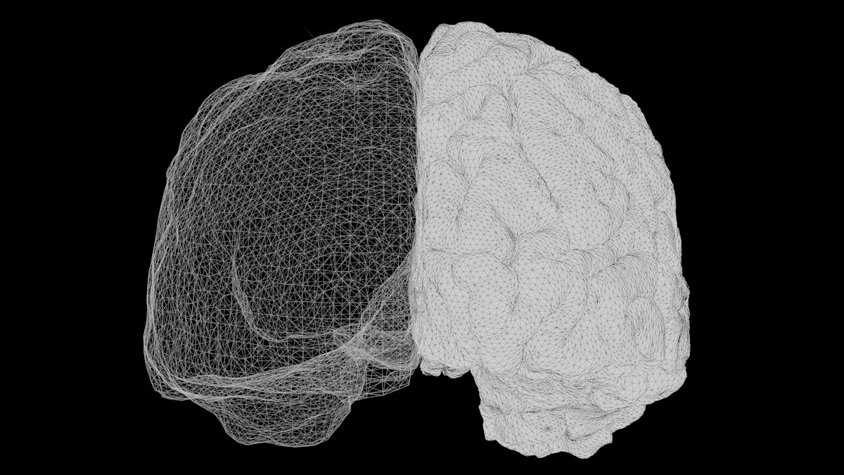
Charles Lieber’s lab at Harvard took that conclusion seriously enough to rebuild the electrode from the ground up. Lieber, a chemist who had spent years working on nanowire and nanotube electronics, started asking what a neural probe would look like if it were designed to match brain tissue mechanically rather than to maximize electrode density. The answer, published in Nature Nanotechnology in 2015 with lead author Jia Liu, was a mesh: a macroporous, open-lattice scaffold of conductive polymer and metal interconnects, so thin and so floppy that it behaves less like a dagger and more like a piece of wet tissue paper.
The brain doesn’t reject the mesh the way it rejects a needle, because the mesh never behaves like a needle.
A Needle, Not a Drill: How the Delivery Actually Works
The trick that makes mesh electronics usable at all is the delivery method. A rigid probe has to be surgically placed, which means drilling, which means a craniotomy sized to the implant. Lieber’s mesh, because it’s thin and flexible enough to fold, can be rolled up and loaded into a glass syringe needle a few hundred microns wide. Injected through that needle into a target region, in the Harvard group’s demonstrations into the brains of live mice, the mesh unfurls once it leaves the needle and settles into the surrounding tissue, free of the syringe, free of any rigid shaft left behind. The entry wound is a needle track, not a surgical opening.
That distinction matters for two separate reasons, one mechanical and one clinical. Mechanically, a structure with a bending stiffness close to that of neural tissue doesn’t fight the brain’s natural movement, so it doesn’t generate the chronic micro-injury that triggers scarring. Clinically, injection instead of craniotomy changes what kind of procedure this could eventually be. A Utah array implant is major neurosurgery. An injected mesh, at least in principle, starts to look like something closer to a lumbar puncture or a deep-brain stimulation lead placement done through a small bore needle rather than an open skull flap.
What the Mouse Data Actually Shows, and What It Doesn’t Yet
The Lieber group’s follow-up work backed the concept with tissue staining, not just electrical recordings. Using GFAP staining to visualize astrocyte activation and Iba1 staining for microglia, the Harvard team found dramatically reduced glial scarring around the mesh compared to rigid silicon probes implanted in the same brain regions, with recordings remaining stable across months rather than the weeks typical of stiff electrode failure curves. Individual neurons could be tracked recording after recording, something that is difficult to do with electrodes that are slowly being walled off by scar tissue.
Twenty years into rigid brain electrodes, the fix wasn’t a better electrode. It was an electrode that stopped acting like a tool.
None of that is a human result. It’s worth saying plainly, because the coverage of this work over the past decade has sometimes blurred the line between “demonstrated in a mouse cortex” and “ready for a patient.” The mesh electronics field is still largely a rodent-stage science. Scaling the injection and unfurling process reliably to a human brain, which is roughly a thousand times larger than a mouse brain and subject to far more stringent regulatory scrutiny, is an open engineering problem, not a solved one. Getting consistent electrode placement, verifying long-term biocompatibility over years rather than months, and building the surgical tooling to do this safely in a human operating room are all real gaps between where the science sits now and where a clinical trial would need it to be.
The Rest of Soft Bioelectronics Is Chasing the Same Insight
Lieber’s mesh is the most dramatic version of a broader shift that’s been happening across neural engineering labs for the past decade. Polina Anikeeva’s group at MIT has built flexible, fiber-based neural probes using thermal drawing techniques borrowed from optical fiber manufacturing, aiming at the same mechanical mismatch problem from a different materials angle. George Malliaras, now at Cambridge, has spent years developing organic electrochemical transistors and conducting polymer coatings that soften the electrode-tissue interface even when the underlying substrate isn’t fully flexible. Zhenan Bao’s lab at Stanford has pushed stretchable, skin-like electronics that borrow some of the same design logic, even where the application is peripheral rather than cortical.
The throughline across all of this work is a bet that the next real gain in chronic neural recording won’t come from packing more channels onto a rigid chip. It will come from making the electrode disappear mechanically into the tissue it’s supposed to be reading. That is a quieter kind of innovation than a new chip architecture, and it has been correspondingly slower to attract headlines, but it may matter more for the long arc of implantable neurotechnology than any single increase in channel count.
Where This Sits Against the Rest of the Field
| Approach | Mechanical Profile | Delivery | Stage |
|---|---|---|---|
| Utah Array (Blackrock Neurotech) | Rigid silicon, gigapascal stiffness | Open craniotomy | Human trials (BrainGate and related consortia) since the 1990s |
| Syringe-injectable mesh electronics (Lieber lab, Harvard) | Tissue-matched flexibility | Needle injection | Rodent studies |
| Stentrode (Synchron) | Semi-flexible metal mesh | Endovascular catheter, no craniotomy | Early human trials |
| Thread electrodes (Neuralink) | Flexible polymer threads, rigid insertion needles | Robotic surgical insertion | Early human implants |
Tom Oxley’s Synchron and Neuralink have both attacked the scarring and surgical-burden problem from different angles than Lieber’s lab. Synchron’s stentrode avoids open brain surgery entirely by delivering a mesh-like electrode array through the vasculature, reading signals from inside a blood vessel pressed against the motor cortex rather than from tissue itself, trading recording resolution for a dramatically less invasive procedure. Neuralink’s threads are flexible once placed but still require a rigid needle and a robotic sewing machine to get them into tissue precisely, which keeps the insertion event itself closer to conventional neurosurgery even though the resulting implant is soft. Mesh electronics, if it scales, offers something none of these fully deliver yet: an implant that is soft at every stage, from the needle that delivers it to the structure that stays behind.
What Changes If Implants Stop Degrading
The philosophical stakes here are easy to understate because the material science sounds so narrow. But think about what chronic signal stability actually enables. Every BCI trial result you’ve read about, the cursor control, the robotic arm, the synthesized speech from motor cortex signals, has been achieved despite a slow electrode failure clock running in the background. Patients in long-running BCI studies have had to recalibrate decoding algorithms as electrode yield dropped, or have had arrays explanted and replaced, or have simply watched performance decline as scar tissue thickened around the implant that gave them a voice or a hand again.
An interface that doesn’t provoke that response changes the shape of the question entirely. It stops being “how long can we keep this working” and starts being “what do we build once it just keeps working.” A neural interface that remains stable for a decade rather than degrading within two or three years is not a faster version of the same technology. It’s a different category of relationship between a nervous system and a machine, closer to a permanent extension than a medical device with a service life.
That reframing deserves some skepticism too. Soft, biocompatible, stable for years: those are exactly the properties regulators and ethicists will scrutinize hardest, because a device meant to stay in a human brain for a decade carries a different risk profile than one meant to be replaced on a predictable schedule. Long-term biocompatibility claims built on months of mouse data need the kind of multi-year, large-animal, then human safety record that no mesh electronics group has yet produced. The honest version of this story is that the mechanical insight is sound and the translational path is long.
What’s true regardless of how fast that path moves is that the field has identified the actual constraint. For twenty years, chronic neural recording was treated as a signal processing problem: more electrodes, better decoding, smarter algorithms compensating for noisy data. Lieber’s mesh work reframes it as a materials problem, one with an answer that was sitting in plain sight the whole time. Make the electrode behave like the tissue around it, and the tissue stops fighting back.