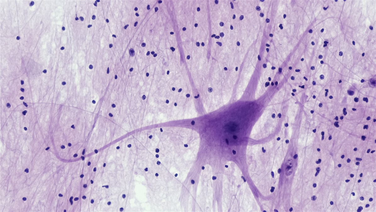
In 1939, at the Marine Biological Laboratory in Plymouth, England, Alan Hodgkin and Andrew Huxley threaded a glass electrode down the inside of a squid giant axon, a nerve fiber wide enough to see with the naked eye, and recorded something no one had measured directly before: a nerve cell firing in real time. The voltage across the membrane didn’t just shift a little when the axon fired. It flipped, swinging from about negative 70 millivolts to positive 40 in under two milliseconds, then snapped back. Hodgkin and Huxley spent the next thirteen years turning that recording into equations precise enough to predict how a nerve impulse behaves before it happens. They won the Nobel Prize in 1963 for it, and the equations are still the starting point for every computational model of a firing neuron built today.
That flip is the action potential. It’s the electrical signal that runs your nervous system, fires your heartbeat, and contracts every muscle in your body. You’ve probably heard it described as an electrical impulse traveling down a wire, and that description is close enough to be useful and wrong enough to be worth fixing. Understanding what actually happens at the membrane, ion by ion, is the difference between reading a metaphor and reading the machine.
A cell keeps its inside charged like a tiny battery, on purpose
Every living cell in your body maintains a voltage difference across its outer membrane, called the membrane potential, and in a resting neuron that difference sits around negative 70 millivolts, inside relative to outside. That number isn’t an accident of biology. It’s built and maintained continuously by a protein called the sodium-potassium pump, which sits in the membrane and spends ATP, your cell’s energy currency, to shove three sodium ions out for every two potassium ions it lets in. The pump runs constantly, all day, in every one of your roughly 86 billion neurons, and it’s the reason a neuron at rest is not electrically neutral but electrically loaded, like a battery sitting on a shelf with charge stored and ready.
The membrane itself is a fatty barrier that ions can’t cross on their own. What lets them through are ion channels, specialized proteins that form pores and open or close depending on the voltage across the membrane. This is the part that makes the whole system work: the channels are voltage-gated, meaning the electrical state of the membrane controls whether the channels open, and the channels opening changes the electrical state of the membrane. It’s a feedback loop built out of protein, and it’s the entire mechanism behind the signal.
The channels open because of voltage, and opening them changes the voltage. That loop is the whole trick.
Why the signal only goes one way, and why that matters more than the spike itself
When a stimulus, whether that’s a synapse firing onto a neuron’s dendrite or a sensory receptor in your fingertip detecting pressure, depolarizes the membrane enough to cross a threshold, usually around negative 55 millivolts, voltage-gated sodium channels along the axon snap open. Sodium ions flood in, because there’s far more sodium outside the cell than inside, and the membrane voltage rockets upward, past zero, up to roughly positive 40 millivolts. That’s the upstroke of the spike. A fraction of a millisecond later, the sodium channels inactivate on their own, a built-in shutoff, and voltage-gated potassium channels open instead, letting potassium rush out and drive the voltage back down, often overshooting slightly below resting potential before the sodium-potassium pump and leak channels restore the baseline.
Here’s the detail that actually explains why nerves work as a communication system and not just a local flicker: right after a patch of membrane fires, it enters a refractory period during which its sodium channels are inactivated and cannot reopen no matter how strong the incoming signal is. That’s what forces the action potential to propagate in one direction down the axon instead of sloshing back and forth. The patch of membrane just behind the spike is temporarily unfireable, so the wave of depolarization only has one direction to go: forward, toward the axon terminal. It’s not that the electricity is directional the way current in a wire is directional. It’s that the membrane makes itself directional, one refractory patch at a time.
The signal doesn’t move because it’s pushed. It moves because the membrane behind it briefly can’t fire again.
This is not electricity the way your house wiring is electricity
Here’s the part worth being direct about, because the wire analogy oversells the similarity. In a copper wire, electrons themselves travel down the conductor, and current moves at a meaningful fraction of the speed of light. In an axon, no single ion travels the length of the fiber. Sodium ions that rush in at one point in the membrane stay roughly where they entered. What propagates is the change in voltage, a wave of channels opening in sequence, like a line of dominoes falling rather than a single domino sliding the whole distance. That’s why nerve conduction, even in the fastest myelinated human axons, tops out around 100 meters per second, which sounds fast until you compare it to electrons in a wire and realize it’s slower by roughly six orders of magnitude.
Myelin is the fix biology found for that speed problem. Oligodendrocytes in the central nervous system and Schwann cells in the peripheral nervous system wrap axons in fatty insulation with periodic gaps called nodes of Ranvier, and because the axon membrane is only exposed to fluid at those nodes, the action potential can effectively jump from node to node rather than regenerating continuously along every micrometer of membrane. This is called saltatory conduction, from the Latin for “leaping,” and it’s the reason a signal from your spinal cord can reach a muscle in your foot in a matter of milliseconds instead of the better part of a second. Multiple sclerosis and Guillain-Barre syndrome are both, at the mechanistic level, myelin-stripping diseases, and the reason they cause weakness and numbness is that the leap gets slower or fails outright, not that the neurons themselves stop working.
The signal has to cross a gap it can’t conduct across
An action potential traveling down an axon eventually hits the axon terminal, and there it runs into a problem: the next cell in the circuit, whether it’s another neuron or a muscle fiber, is separated by a synaptic cleft, a gap of about 20 nanometers that the electrical signal cannot jump on its own. Biology’s answer is to convert the electrical signal into a chemical one and back again. The arriving action potential opens voltage-gated calcium channels at the terminal, calcium flooding in triggers vesicles full of neurotransmitter, commonly glutamate, GABA, acetylcholine, or dopamine depending on the circuit, to fuse with the membrane and dump their contents into the cleft. Those neurotransmitter molecules diffuse across the gap in fractions of a millisecond and bind receptors on the receiving cell, which in turn opens ion channels there and either pushes that cell’s membrane toward its own firing threshold or away from it.
This is the point where drugs and diseases intervene, because the synapse is a chemical bottleneck in an otherwise electrical system, and chemistry is easier to manipulate with a pill than voltage is. SSRIs work by blocking the reuptake of serotonin at the synapse. Botulinum toxin works by blocking vesicle fusion at the neuromuscular junction, which is why it paralyzes muscle. Myasthenia gravis is an autoimmune attack on acetylcholine receptors at that same junction. None of these interventions touch the action potential itself. They all work on the five-to-twenty-nanometer chemical handoff between spikes.
What clinicians actually do with this, and where it gets exploited
Nearly every piece of modern neurology and cardiology hardware that reads or writes to the body is built on the fact that action potentials are electrical events you can detect from outside the cell. An EEG picks up the summed electrical activity of thousands of cortical neurons firing together. An EKG reads the coordinated action potentials of cardiac muscle cells as they trigger each heartbeat, and a pacemaker works by directly injecting a small current to force cardiac cells past threshold when the heart’s own pacemaker cells fail to. Deep brain stimulation electrodes, used in Parkinson’s disease since the FDA approved the first system in 1997, deliver current directly to basal ganglia structures to override pathological firing patterns. Cochlear implants, approved by the FDA in 1984, bypass damaged hair cells entirely and trigger action potentials directly in the auditory nerve using electrode arrays tuned to different sound frequencies.
| Device or condition | What it reads or writes | Relies on |
|---|---|---|
| EEG | Summed cortical activity, read only | Extracellular voltage from synchronized firing |
| Cardiac pacemaker | Heart muscle, read and write | Forcing cardiac cells past threshold |
| Deep brain stimulation | Basal ganglia, write only | Overriding pathological firing patterns |
| Cochlear implant | Auditory nerve, write only | Directly triggering action potentials by frequency |
| Local anesthetic (lidocaine) | Peripheral nerve, block only | Physically plugging sodium channels shut |
Local anesthesia is worth a mention here because it shows the mechanism from the opposite direction. Lidocaine and its relatives work by physically wedging themselves into voltage-gated sodium channels and jamming them shut, which stops action potentials from firing at all in the treated tissue. No sodium channels opening means no upstroke, no threshold crossing, no pain signal reaching your spinal cord. It’s the cleanest possible demonstration that the entire elaborate signaling system depends on one specific class of protein doing its job, and blocking that one protein is enough to shut the whole channel of communication down, locally and reversibly.
Where the tidy model runs into real limits
The Hodgkin-Huxley picture is clean, and that’s exactly the reason to be careful with it. It describes a squid axon, a single fiber optimized by evolution for speed and simplicity, and human neurons are messier. Cortical neurons carry a mix of channel types that Hodgkin and Huxley never measured, dendrites can generate their own local spikes that never make it to the main axon, and glial cells, long dismissed as passive support tissue, are now known to modulate neuronal firing in ways the original model doesn’t account for at all. Treat the classic action potential model as a foundation, not a complete map of what a neuron does. It explains the spike. It doesn’t fully explain the neuron.
What the model does explain, precisely and reliably, is why bioelectronic medicine works at all. Every implant that talks to your nervous system, from a cochlear implant to an experimental spinal cord stimulator restoring movement in paralyzed patients, as Gregoire Courtine’s group at EPFL has demonstrated, is exploiting the same negative 70 millivolt battery and the same voltage-gated channels that Hodgkin and Huxley described from a squid on a dissection tray in Plymouth in 1939. The squid axon was never the point. It was the simplest possible version of a mechanism your body runs, in parallel, in trillions of copies, every second you’re alive.